3264
Investigation of Cerebral Structural Alterations of Patients with Mild Cognitive Impairment based on VBM and DBM Analysis1Capital Medical University, Beijing, China, 2Clincial Science, Philips Healthcare, Guangzhou, China
Synopsis
Voxel-based morphometry (VBM) and deformation-based morphometry (DBM) are wildly-used automated analysis pipelines for neuro-structural images. This study aims to investigate gray matter abnormalities in mild cognitive impairment (MCI) patients. Some altered brain regions could be detected with VBM and DBM method for MCI patients compared to healthy controls. Besides, the features extracted from VBM and DBM could effectively identify the MCI from healthy controls.
Introduction
Mild cognitive impairment (MCI) is a brain function syndrome which causes a slight but noticeable and measurable decline in cognitive abilities. A person with MCI is at an increased risk of developing to Alzheimer's Disease. So it is necessary and important to explore reliable biomarkers for clinical MCI early detection. The aim of this study was to investigate the structural biomarkers of MCI by using VBM and DBM method1,2.Methods
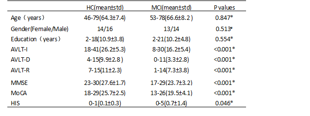
27 MCI and 30 age-, sex- and education-matched healthy controls (HC) were included in this study. Each subject signed an informed consent form and this study was approved by the ethics committee of Capital Medical University Xuanwu Hosipital. MR images were obtained by using a high-resolution 3D T1-weighted sequence with the following parameters: Echo Time(TE) / Repetition Time(TR) = 2.2ms / 1900ms, Matrix=448×512 and Voxel Size =111mm3. Firstly, the structural MRI data were analyzed by VBM and DBM using CAT12 software (refer to the CAT12 manual for details of the processing steps). Then, multiple comparison two-sample t tests (p<0.05, Cluster Size>178 voxels for VBM, Cluster Size>113 voxels for DBM) were performed on the processed images to detect the difference of MCI and HC. Furtherly, a support vector machine based classification method was adopted to identify MCI patients from HC using the features extracted from the altered brain regions3.Results
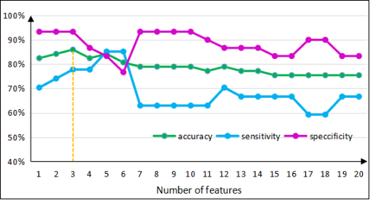
Details of demographics and clinical characteristics are presented in Table 1. Fig.1 shows the results of VBM and DBM analysis. The two methods both showed that the significantly changed brain regions mainly included the bilateral para-hippocampal, hippocampus, amygdala, insula and inferior frontal gyrus. In addition, the VBM analysis also displayed changes primarily laid in the bilateral superior frontal gyrus, middle frontal gyrus, middle temporal gyrus, inferior temporal gyrus, superior occipital gyrus, middle occipital gyrus, supplementary motor area and right cingulum gyrus. The SVM classification accuracies were 71.9% and 77.2% for VBM and DBM, respectively. Fig.2 shows the classification accuracy versus the number of features extracted from VBM results. The best accuracy of 86% was obtained with the optimized features selection by Fisher Score. Table 2 lists the accuracy, sensitivity and specificity for the classification in detail.Discussion
Gray matter abnormalities in MCI patients could be detected with VBM and DBM methods. VBM detects altered brain regions mainly via evaluating the changes of gray matter volume, while DBM reflects alterations primarily by the value of Jacobian determinant produced during registration process. Our study shows that, for MCI patients, gray matter changes mainly locate in para-hippocampal, hippocampus, amygdala and inferior frontal gyrus that are relevant to cognitive functions. Furthermore, we did feature selection for VBM extracted features by Fisher Score algorithm and significantly improved the classification performance. DBM method could effectively find the more discriminative characteristics according to the classification results. The results are helpful for the recognition of MCI in clinical practice.Conclusion
VBM and DBM methods both could detect some alterated brain regions for MCI patients compared to healthy controls. Besides, the features extracted from VBM and DBM could effectively identify the MCI from healthy controls.Acknowledgements
No acknowledgement found.References
1. Zhang W, Song L, Yin X, et al. Grey Matter Abnormalities in untreated hyperthyroidism: a voxel-based morphometry study using the DARTEL approach [J]. European Journal of Radiology, 2014, 83(1): 43-48.
2. Savio A, Grana M. Deformation based feature selection for computer aided diagnosis of alzheimer’s disease[J]. Expert Systems with Applications, 2013, 40(5): 1619-1628.
3. Magnin B, Mesrob L, Kinkingnéhun S, et al. Support vector machine-based classification of Alzheimer’s disease from whole-brain anatomical MRI [J]. Neuroradiology, 2009, 51(2): 73-83.
Figures