3250
A 3D-printed anatomical multimodal phantom for brain segmentation validation1Department of Biomedical Engineering, University of Basel, Allschwil, Switzerland, 2Medical Image Analysis Center (MIAC) AG, Basel, Switzerland, 3Department of Radiology, University Hospital of Basel, Basel, Switzerland, 4Department of Neurology, University Hospital of Basel, Basel, Switzerland, 5DKD HELIOS Klinik, Wiesbaden, Germany
Synopsis
Brain tissue segmentation algorithms applied on magnetic resonance imaging (MRI) data lack a ground truth for evaluating their performance. For this purpose, an anatomical brain phantom prototype mimicking T1 relaxation times and the complex 3D geometry of the human brain was created for use with MRI and computed tomography (CT). A scan-rescan experiment showed a low within-session variability of white matter (WM) and grey matter (GM) volumes when MRI images of the phantom were segmented with a commonly used software. Compared to the ground truth volumes derived from CT, the software overestimated the WM, while the GM was slightly underestimated.
Purpose
Quantification of brain tissue volumes in magnetic resonance imaging (MRI) is essential when assessing and monitoring neurodegenerative processes. However, the precise quantification largely depends on the image acquisition or processing as well as on the performance of the applied tissue segmentation tools [1]. In this regard, a ground truth to disentangle confounding methodological factors (e.g. scanner, imaging protocol, software features) from volume fluctuations due to biological and pathological changes is still missing. We here propose the use of a human brain phantom mimicking grey (GM) and white matter (WM) structure as a ground truth for evaluating the accuracy and reliability of a commonly used segmentation software.Methods
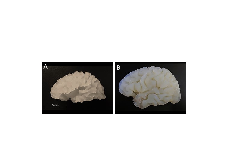
The brain phantom was built according to procedures recently described [2]. Briefly, the samples reproducing the T1 relaxation times of WM and GM were prepared with, respectively, 0.12 mM and 0.04 mM of MRI contrast agent (manganese chloride, MnCl2) diluted in a 0.6% agar solution. 24 mg/mL of the computed tomography (CT) contrast medium Iopromidium was added to the WM sample. The complex 3D geometry of the two compartments was based on WM and GM surfaces automatically derived from a healthy subject MPRAGE MRI scan (resolution = 1x1x1 mm3, TR/TI/TE = 1570/900/2.48 ms, α = 8, 3T scanner, 20-channel head coil). Those surfaces were then 3D-printed using polylactic acid thermoplastic and covered by a brushable platinum-cure silicone rubber to obtain flexible molds. Finally, the different parts of the phantom were assembled: the WM sample was injected into the WM surface mold; once the sample gelled, the mold was removed, a hydrophobic varnish layer was applied and the GM mold was positioned on top; then, the GM sample was injected and the corresponding mold was pulled off when the gel became solid; finally, a silicone shell mimicking the skull was positioned on top of the gel composite.
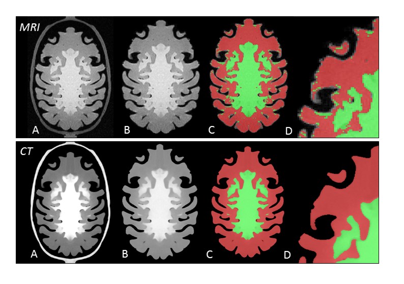
Once the phantom was assembled, ground truth volumes were calculated from CT images (resolution = 0.44x0.44x0.25 mm3, 120 kV, 270 mAs, J30 reconstruction kernel, 226 mm FOV). After a manual brain extraction, the intensity distribution of the phantom brain was shifted by 1000 HU and surrounding noise was removed by applying a threshold (t = 850 HU). All the voxels characterized by an intensity ≥ 1160 HU were recognized as WM, while voxels with intensity < 1160 HU were assigned to GM. The ground truth volumes were computed as the number of voxels in each class multiplied by the voxel dimensions. To assess the accuracy of the segmentation software, MRI images of the phantom were acquired using the same MPRAGE sequence used for the healthy volunteer scan (see above). From these images, the phantom skull was removed using BET (FSL, [3]) and the remaining phantom brain was segmented using FAST (FSL, [4]). WM and GM volumes were computed from the resulting segmentation by multiplying the voxel numbers assigned to WM or GM by the voxel dimensions. The reliability of FAST was further assessed through a scan-rescan test. Since the phantom only reproduces one hemisphere, all acquired images were mirrored at the mid-sagittal plane to simulate a whole brain.
Results
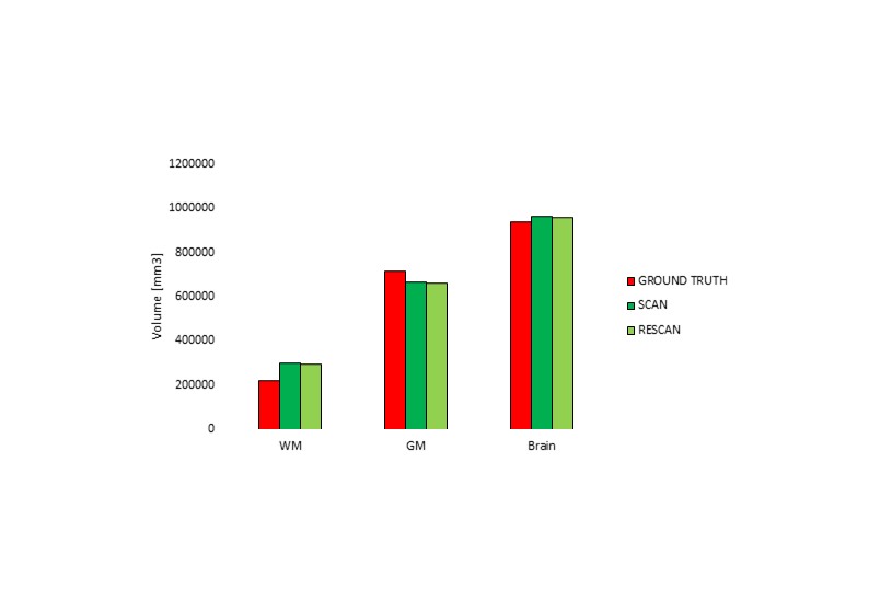
The phantom with plausible and homogenous T1 contrast as well as realistic structure was successfully built (Fig. 1, 2). The difference between scan-rescan in the computed volumes was less than 1% (Brain: 0.81%; WM: 0.79%; GM: 0.81%, Fig. 3). The ground truth volume assessed by CT was 942490 mm3 for the whole brain, 224890 mm3 for the WM, and 718009 mm3 for the GM compartments. The segmentation performed by FAST showed a difference of +2.73% for the total brain volume, an overestimation of the WM by +34.06%, and an underestimation of the GM equal to -7.13% (Fig. 3). The voxels wrongly classified as WM instead of GM were mostly located at the WM/GM interface as well as at the outer border of the GM (Fig. 2).Discussion
Our results indicate that FAST is characterized by a low scan-rescan variability. Moreover, whole-brain volumes derived from FAST are very close to the ground truth. WM and GM volumes differ from the ground truth, especially in the case of WM. This finding could be partially attributed to some voxels in the outer part of the GM affected by susceptibility artefacts and wrongly assigned to the WM. Filling of the cerebrospinal fluid compartment with water will likely allow to mitigate this effect.Conclusions
Our brain phantom, with its structural and intensity properties, allows the testing of automatic segmentation software like FAST. Further phantom developments will allow to even more realistically simulate the performance of brain segmentation.Acknowledgements
No acknowledgement found.References
[1] Despotovic, I., Goossens, B., Philips, W., 2015. MRI Segmentation of the Human Brain: Challenges, Methods, and Applications 2015.
[2] Altermatt, A., Santini, F., Deligianni, X., Magon, S., Cattin, P., Wuerfel, J., Gaetano, L., 2017. Novel Design of a 3D-Printed Anthropomorphic Brain Phantom for Segmentation Validation in Magnetic Resonance Imaging. ISMRM Annu. Meet.
[3] Smith, S.M., 2002. Fast Robust Automated Brain Extraction. Hum. Brain Mapp. 17, 143–155. doi:10.1002/hbm.10062
[4] Zhang, Y., Brady, M., Smith, S., 2001. Segmentation of brain MR images through a hidden Markov random field model and the expectation-maximization algorithm. IEEE Trans. Med. Imaging 20, 45–57. doi:10.1109/42.906424
Figures