3218
L-spine Bone Marrow on Female: A Intravoxel Incoherent Motion MR Imaging Study1Department of Medical Imaging, Affiliated Hospital of Shaanxi University of Traditional Chinese Medicine, Xianyang, China, 2Department of Medical Imaging, The First Affiliated Hospital of Xi'an Jiaotong University, Xi'an, China
Synopsis
To our knowledge, no studies have employed IVIM diffusion-weighted MRI to explore the variation trend of bone marrow in female. Whether gender difference exists in marrow structure is not well investigated. Therefore, we explored the diagnostic utility of IVIM diffusion-weighted MRI parameters in this context. We found that D, D* and f value showed a decreased trend with age, and the D, D* value of bone marrow in female was significantly higher than that in male except the f value. IVIM diffusion-weighted MRI was useful in the evaluation of bone marrow.
Introduction:
Female's estrogen and menstrual cycle can affect the conversion of bone marrow, which induce the changes of bone metabolism, bone mass and bone microstructure. Knowledge of the normal MR appearance of marrow conversion with age is necessary for the recognition of abnormal conversion and reconversion patterns as well as infiltration of marrow by tumor and other pathologic processes. Intravoxel Incoherent Motion (IVIM) diffusion-weighted MRI is increasingly employed clinically to evaluate tissue perfusion without the use of contrast agents. The ability to analyze non-Gaussian diffusion via high-diffusion weighting enhances the detection sensitivity of tissue features. This study was designed to explore the changes of female bone marrow based on IVIM technology.Methods:
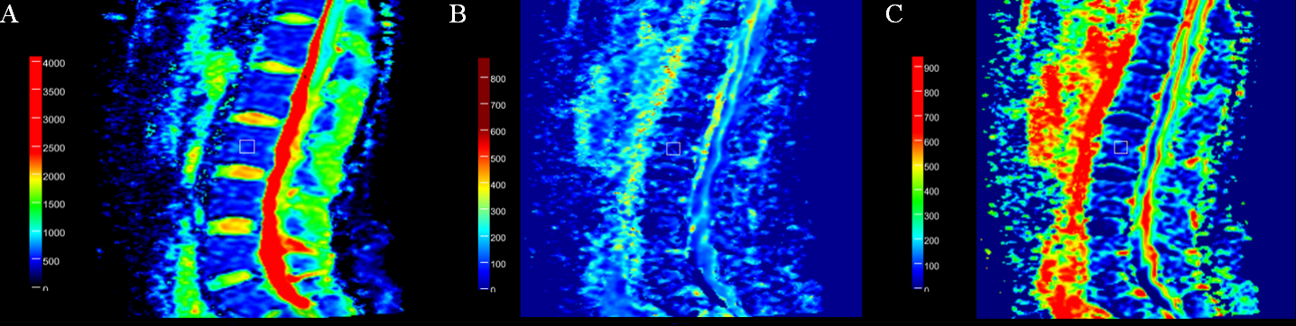
Sixty-four healthy adult subjects (36 females and 28males) were involved in this study, who were divided into youth group ( < 40 years), middle-aged group ( 40~59 years), elderly group( > 59 years). The parameters of the five lumbar vertebrae among each subject were measured by a prototype diffusion post-processing tool called MR body Diffusion Toolbox1.0, which measured a total of 320 vertebral body. The MRI scans were acquired with the use of 3T MRI scanner (MAGNETOM Skyra, Siemens healthcare, Erlangen, Germany), including collected T1-weighted, T2-weighted and IVIM sequences. IVIM sequence was performed with 8 b values (b=0, 50, 100, 150, 200, 400, 600 and 800 s/mm2), repetition time = 2800ms, echo time = 75ms, field of view = 220x220 mm2, matrix = 83×118, slice thickness = 4mm, gap = 1.2mm. The molecular diffusion coefficient (D), the perfusion-related D (D*), and perfusion fraction (f) were calculated by the IVIM bi-exponential model. All parameters were compared between different gender groups and among age groups. The association between skeletal system D value, D* value, f value and age was also investigated using Pearson’s correlation respectively.Results:
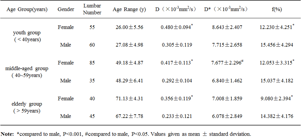
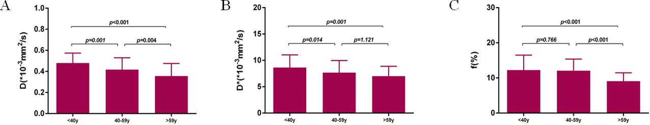
The D, D* value of female were higher than that of male, while f value was lower in female in each age group compared with male (Table 1). All D, D* and f value showed a decreased trend with age in different age group of female (Figure 2). Negative correlations were observed between skeletal system D, D*, f values and their age in female respectively.Discussion:
The female showed a decreasing trend of D value with age, which might be caused by bone marrow structure conversion. The D * value was significantly reduced in female when their age was older than 40 years, suggesting that the average diameter of bone marrow capillary and average blood flow rate decreased. The f value which indicates perfusion was significantly reduced in female in elderly group. Our current findings indicated that there was a significant difference of D* value in gender in middle-aged group, while this difference between the youth group and the elderly group is not obvious. The difference might result from that the female menstrual cycle and estrogen effects which stimulate bone marrow hematopoietic cell proliferation and promote red bone marrow formation. The D value in female was higher than that in male. However, the f value in female was lower than male. IVIM parameters decreased gradually with age. There was a negative correlation between the parameters on IVIM and age in female.CONCLUSION:
There were significant differences in IVIM parameter values between female and male. Furthermore there was a significant downward trend with age in female. According to the changes of D, D *, f value, IVIM can be used as a monitoring means of female bone marrow transformation process.Acknowledgements
Author contributions: Study design: Y.-s. Zheng, T. Zhao, N. Yu. Definition of intellectual content: T. Zhao. Literature search: Y.-y. Chen, Y.-b. Guo, T. Zhao. Data acquisition: Y.-y. Chen, Y.-b. Guo and T. Zhao. Data analysis: T. Zhao and D. Han. Statistical analysis: T. Zhao and D. Han. Manuscript editing: T. Zhao, H. Xu, Y.-s. Zheng. Manuscript review: H. Xu, Y.-s. Zheng.References
1. Bourillon C, Rahmouni A, Lin C, et al. Intravoxel Incoherent Motion Diffusion-weighted Imaging of Multiple Myeloma Lesions: Correlation with Whole-Body Dynamic Contrast Agent-enhanced MR Imaging. Radiology, 2015, 277(3):773-783.
2. He J, Fang H, Na L X. Vertebral bone marrow diffusivity in normal adults with varying bone densities at 3T diffusion-weighted imaging. Acta Radiologica, 2017:284185117704235.
3. Ohno N, Miyati T, Kasai H, et al. Evaluation of perfusion-related and true diffusion in vertebral bone marrow: a preliminary study. Radiological Physics & Technology, 2015, 8(1):135-140.
Figures