3216
Diffusion kurtosis imaging in evaluating non-Hodgkin’s lymphoma: associations with tumor aggressiveness and proliferation1Radiology, Fujian Cancer Hospital, Fuzhou, China, 2Philips Healthcare, Shanghai, China
Synopsis
This study tries to evaluate the usefulness of diffusion kurtosis imaging (DKI) parameters in segregating the pathological subtypes of NHL lymphoma, and to explore its associations with aggressiveness and proliferative index. Significant differences were found among the WHO classified subgroups. Meanwhile, the stronger tumor aggressiveness (higher Ki67 percentage) was accompanied with more restricted water diffusivity (lower ADC and D values) and more complex cell micro-structural environment (higher K value). The DKI technique may help estimation of tumor proliferation in NHL lymphoma and DKI parameters can be used as imaging biomarker of the biological aggressiveness of the tumor.
Introduction & Purpose
Non-Hodgkin’s lymphoma is a disease both clinically and biologically heterogeneous encompassing divergent tumor types with contrasting clinical behaviors. Traditional grading methods like nuclear pleomorphism and mitotic count do not apply to estimate the invasiveness and proliferation of lymphoid neoplasms. This study aims to evaluate the usefulness of diffusion kurtosis imaging (DKI) related parameters in segregating the pathological subtypes of lymphoma, and to explore its associations with aggressiveness and proliferative index.Methods
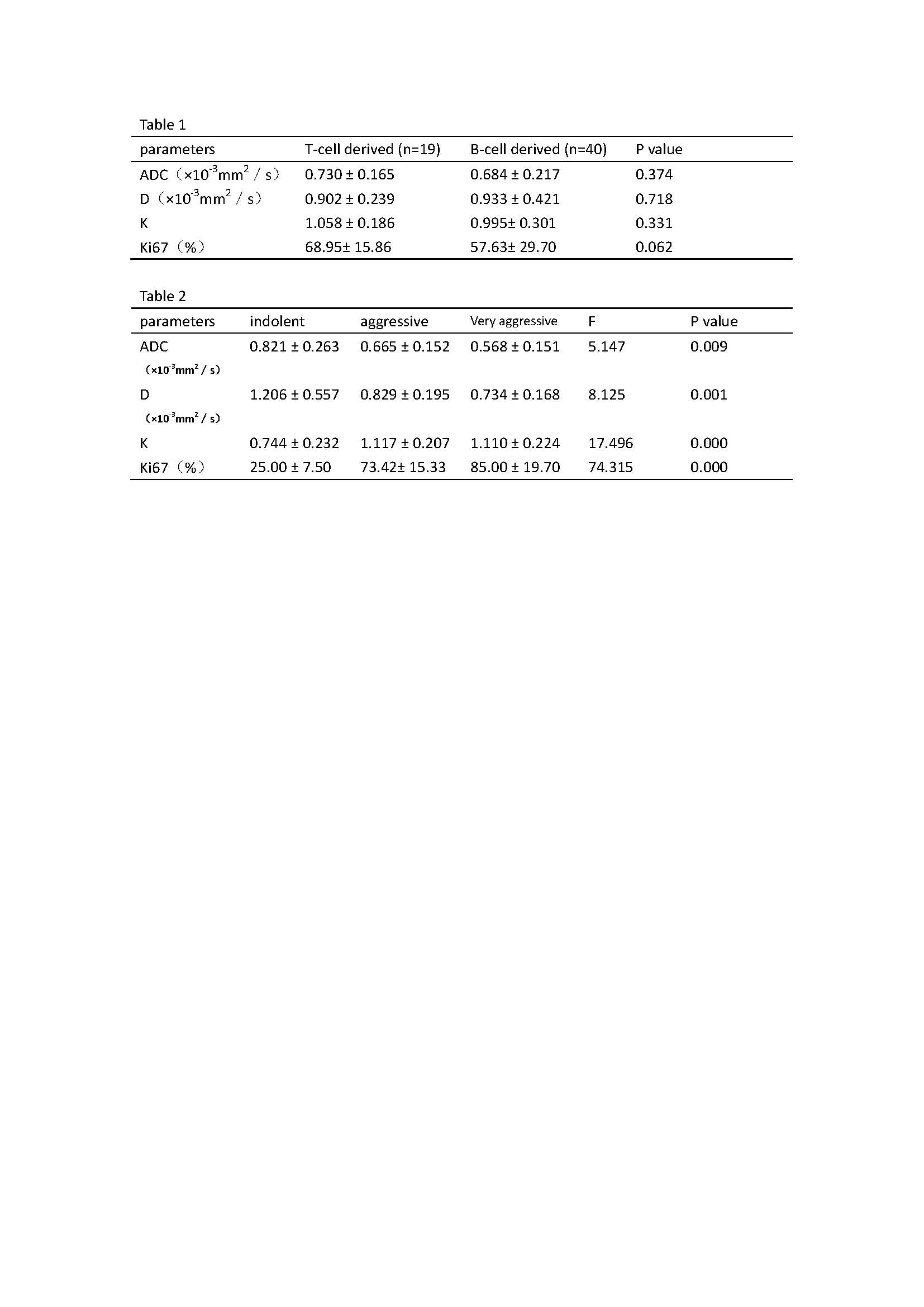
59 consecutive patients with newly diagnosed lymphoma (range, 20-86 years, median, 53 years, female/male 11/48) underwent pretreatment DKI examinations on 3T MR scanner with a 16-channel head and neck array coil. Acquisition parameters were: TR/TE 4190/69 ms, matrix size 224 × 224, FOV 240 × 240 mm, slice thickness/gap 5/1 mm, Sense factor 3, NSA 2, b value 0, 500, 1000, 1500, 2000 sec/mm2, scan time 4 min 33s. DKI-related parameters including corrected diffusion coefficient (D) and apparent kurtosis coefficient (K) were calculated. The tumor proliferation was assessed by the immunohistochemical analysis of Ki67 expression. The patients were divided into different subgroups based on pathological type (T cell and B cell derived), WHO classification[1,2] (indolent, aggressive and very aggressive). The DKI parameters of subgroups were compared and the relationship between DKI related parameters and proliferative index were analyzed statistically. Findings with a P value less than 0.05 were considered as statistical significance.Results
The comparisons of DKI-related parameters and proliferative index between pathological subtypes were shown in table 1. Table 2 showed the significant difference of ADC, D, K values and Ki67 percentage among indolent, aggressive and very aggressive lymphoma. The Pearson analysis indicated that either ADC or D value had strongly negative relationship with K value and Ki67 level (ADC/ D to K, r= -0.424/-0.510, P<0.05; ADC/D to Ki67, r= -0.464/- 0.426, P<0.05). In addition, K value was positively correlated with Ki67(r=0.612, P<0.001).Discussion
The DKI-related parameters showed no significant difference between T cell and B cell derived lymphoma, but significant difference among the indolent, aggressive and very aggressive subtypes of lymphoma. Moreover, our findings also suggested that the stronger tumor aggressiveness (higher Ki67 percentage) was accompanied with more restricted water diffusivity (lower ADC and D values) and more complex cell micro-structural environment (higher K value) in NHL lymphoma.Conclusions
The DKI technique may permit estimation of tumor proliferation in NHL lymphoma and DKI parameters can be used as imaging biomarker of the biological aggressiveness of the tumor.Acknowledgements
No acknowledgement foundReferences
[1] Hernández J, Krueger J E, Glatstein E. Classification of Non-Hodgkin's Lymphoma: A Proposal. Oncologist, 1997, 2(4):235.
[2] Jaffe ES, Harris NL, Stein H, Vardiman JW, editors. Pathology and Geneticsof Tumors of Hematopoietic and Lymphoid Tissues. World Health OrganizationClassification of Tumors, Vol. 3. Lyon, France: IARC Press; 2001.
Figures