3213
A Local Coregistration for Quantitative Evaluation of Distortion of Parotid Gland Tumors in Echo-Planar DWI and PROPELLER DWI1Master 's Program of Biomedical Informatics and Biomedical Engineering, Feng Chia University, Taichung City 407, Taiwan, 2Department of Automatic Control Engineering, Feng Chia University, Taichung City 407, Taiwan, 3Ph. D. Program of Electrical and Communications Engineering, Feng Chia University, Taichung City 407, Taiwan, 4Department of Radiology, Tri-Service General Hospital, Taipei, Taiwan, 5Department of Diagnostic Radiology, The University of Hong Kong, Hong Kong, Hong Kong, 6Ph.D. program of Technology Management, Chung Hua University, Hsinchu, Taiwan, 7Department of Medicine, Taipei Medical University, Taipei, Taiwan, 8Department of Dentistry, National Defense Medical Center, Taipei, Taiwan, 9Department of Radiology, National Defense Medical Center, Taipei, Taiwan, 10Master 's Program of Biomedical Informatics and Biomedical Engineering, Feng Chia University, TAICHUNG, Taiwan, 11Institute of Automatic Control Engineering, Feng Chia University, TAICHUNG, Taiwan
Synopsis
The study is to quantitatively compare the morphology distortion in distinguishing parotid pleomorphic adenomas (PMA) between PROPELLER-DWI and EP-DWI. This retrospective study enrolled 14 PMAs. All participants underwent 1.5-T fat-saturated diffusion-weighted imaging with PROPELLER-DWI and EP-DWI. A local coregistration method to quantitatively evaluate the distortion of parotid gland tumors between single shot EP-DWI and PROPELLER DWI. Imaging distortion represented by Dice coefficient was quantitatively analyzed. Our results showed that PROPELLER-DWI allows distinguishing PMAs with less distortion than EP-DWI.
Introduction
In salivary glands, single shot EP-DWI has been used to evaluate the diffusivity of parotid tumors since 2001.1-3 However, single shot EP-DWI is susceptible to artifacts. In head and neck, magnetic susceptibility artifacts and motion artifacts are prominent due to abundant air-soft tissue interface4, metallic implant 4,5, and involuntary bulk motion 6. Compared to single shot EP-DWI , PROPELLER-DWI has also been applied to healthy parotid glands to reduce geographic distortion and chemical shift artifacts in 20094. In this study, we used a local coregistration method to quantitatively evaluate the distortion of parotid gland tumors between single shot EP-DWI and PROPELLER DWI.Materials and Methods
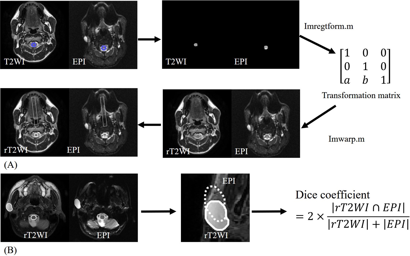
MR scan: A total of 14 patients who had the pleomorphic adenomas (PMA) and received both PROPELLER-DWI and EP-DWI were enrolled. All MR scans were performed at a 1.5 T whole-body scanner (GE Healthcare, Signa HDx, US) using an 8NV head and neck array coil. A T2WI images for coregistration of all participants were scanned by FSE (TR/TE/NEX 3150/78/2, FOV: 250 mm, 512x512, 5 mm thickness). DWI images were obtained with motion-probing diffusion gradients (b factors, 0 and 1000 sec/mm2) applied in each of three orthogonal directions. For PROPELLER-DWI, fast spin-echo sequences (TR/TE/NEX=7000/76/1.8) were performed with fat saturation by using an echo train length of 24, as described by Pipe et al7. For same scan time with PROPELLER-DWI, EP-DWI acquisitions (TR/TE/NEX=7000/73.3/4) were performed without acceleration. Data analysis: All MR data were digitally transferred from the MR unit console to a personal computer and processed with software developed in house by using Matlab (MathWorks, Natick, MA). On T2WI, PROPELLER-DWI and EP-DWI, one slice containing the largest area of the parotid tumor and another slice containing the largest area of contralateral parotid gland were used for quantitative data analysis in each tumor, respectively. T2WI was used to be the reference image for quantitatively evaluating image distortion of PROPELLER-DWI and EP-DWI. The basic idea of our local coregistration method was to find a local object as an origin region and then coregistrate two local objects at same location in two images. Firstly, a ROI contouring cervical spinal cord was defined as the origin region because of its central location, well demarcation, and higher signal intensity than other tissues on DWI. A rigid-body transformation matrix for image registration between T2WI and b0 image of DWI was then calculated by an intensity-based image registration approach. In the next step, the whole image of T2WI was reconstructed from the transformation matrix. Finally, image registration among reconstructed T2WI, b0 image of PROPELLER-DWI, and b0 image of EP-DWI was virtually verified using two ROIs (cervical spinal cord and a rectangular area at the midline of oral cavity and oropharynx), which were drawn on T2WI and then automatically copied to corresponding b0 image of PROPELLER-DWI, and b0 image of EP-DWI (Fig.1A). After image registration, the parotid tumors are manually contoured on rT2WI, PROPELLER-DWI and EPI, respectively. Then two ROIs are overlapped on rT2WI to provide visual perception of image distortion. Dice similarity coefficient is computed finally (Fig.1B)Result
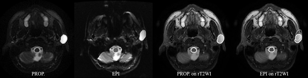
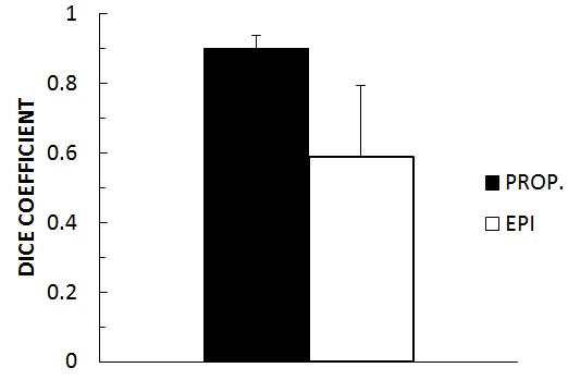
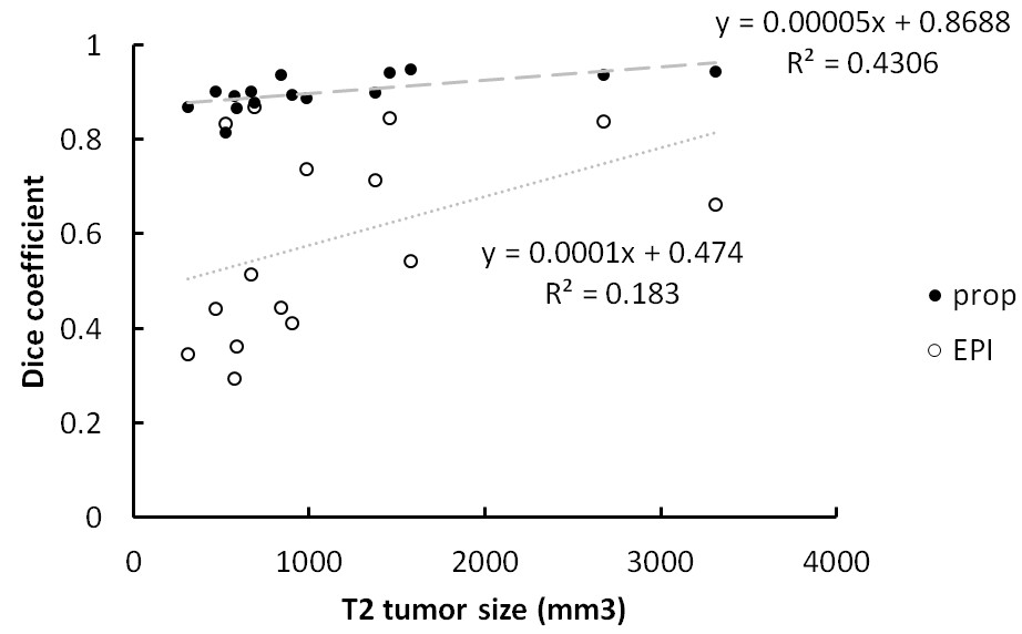
A case demonstration of imaging distortion on PROPLLER-DWI and EP-DWI was shown on Fig. 2. Fig. 3 illustrated Dice coefficients of PROPELLER-DWI and EP-DWI regarding tumor distortion. The Dice coefficients on PROPELLER-DWI VS. rT2WI was 0.897 ± 0.035, and on EP-DWI VS. rT2WI was 0.594 ± 0.212. Scatterplot demonstrating the relationship between the Dice coefficient and ROI size was shown on Fig 4.Discussion
Whether imaging distortion on EP-DWI differs among parotid tumors or not has never been investigated before. The study has quantitatively evaluated the morphology change of parotid tumor between T2WI and DWI using Dice coefficients by the coregistration of spinal cord as origin region. Our results show that the distortion of tumor was slight change on PROPELLER-DWI only, but the geographic distortion on EP-DWI is more severe. The relationship between the tumor size and the severity of distortion indicated it was independent of each other for PROPELLER-DWI, but the distortion of tumor might be affected by the tumor size in EP-DWI.Conclusion
The local coregistration method could provide a quantitative evaluation of geometric distortion of parotid gland tumors in EP-DWI.
Acknowledgements
The study was supported partly from the Ministry of Science and Technology, R. O. C. under the Grant No. MOST 105-2221-E-035 -049 -MY2 and 105-2314-B-016 -024 -MY2.References
1. Wang J, Takashima S, Takayama F, et al. Head and neck lesions: characterization with diffusion-weighted echo-planar MR imaging. Radiology. 2001;220(3):621-30.
2. Eida S, Sumi M, Sakihama N, Takahashi H, Nakamura T. Apparent diffusion coefficient mapping of salivary gland tumors: prediction of the benignancy and malignancy. AJNR Am J Neuroradiol. 2007;28(1):116-21.
3. Habermann CR, Arndt C, Graessner J, et al. Diffusion-weighted echo-planar MR imaging of primary parotid gland tumors: is a prediction of different histologic subtypes possible? AJNR Am J Neuroradiol. 2009;30(3):591-6.
4. Juan CJ, Chang HC, Hsueh CJ, et al. Salivary glands: echo-planar versus PROPELLER Diffusion-weighted MR imaging for assessment of ADCs. Radiology. 2009;253(1):144-52.
5. Chang HC, Juan CJ, Chiu HC, et al. Parotid fat contents in healthy subjects evaluated with iterative decomposition with echo asymmetry and least squares fat-water separation. Radiology. 2013;267(3):918-23.
6. Liu YJ, Lee YH, Chang HC, et al. A potential risk of overestimating apparent diffusion coefficient in parotid glands. PLoS One. 2015;10(4):e0124118.
7. Pipe JG, Farthing VG, Forbes KP. Multishot diffusion-weighted FSE using PROPELLER MRI. Magn Reson Med. 2002;47(1):42-52.
Figures