3210
Improvements in Whole Body Diffusion Weighted Imaging: Combination of Integrated Slice-Specific Dynamic Shimming and Readout-Segmented EPI1Siemens Shenzhen Magnetic Resonance Ltd, Shenzhen, China, 2Siemens Healthcare, Erlangen, Germany
Synopsis
Single-shot echo planar imaging (ss-EPI) is most frequently used for whole body diffusion weighted imaging (WB-DWI) because of short acquisition time and motion insensitivity. However, ss-EPI is vulnerable to the effects of the static field inhomogeneity and poses a challenge to perform ss-EPI based WB-DWI at 3 Tesla. Integrated slice-specific dynamic shimming (iShim) combined with ss-EPI has shown a remarkable improvement on the susceptibility related artifacts in WB-DWI. In this study, we demonstrate the application of rs-EPI using iShim to WB-DWI, which can provide higher quality WB-DWI, specifically less spatial distortions.
INTRODUCTION
Whole body diffusion-weighted imaging (WB-DWI) has been widely used to detect, characterize and monitoring tumors 1. Single-shot echo planar imaging (ss-EPI) is most frequently used for WB-DWI because of short acquisition time and motion insensitivity. However, ss-EPI is vulnerable to the effects of the static field inhomogeneity and poses a challenge to perform ss-EPI based WB-DWI at 3 Tesla. For example, the stronger distortions and signal loss in the neck region of conventional WB-DWI are very common, resulting in a well-known problem (“broken spine”) in reformatted sagittal MPR images 2. Recently the integrated slice-specific dynamic shimming (iShim) combined with ss-EPI was proposed to improve the susceptibility related artifacts and has shown a good performance on WB-DWI 3-4.
Readout-segmented Echo Planar Imaging (rs-EPI) with 2D navigation 5 is an established clinical technique for acquiring diffusion-weighted (DW) images with a low level of distortion and T2*-related blurring. The iShim technique has been extended to rs-EPI and shown improved image quality in the head/neck DW scans, compared with ss-EPI-iShim and conventional rs-EPI 6. In this study, we demonstrate the application of rs-EPI using iShim to WB-DWI.
METHODS
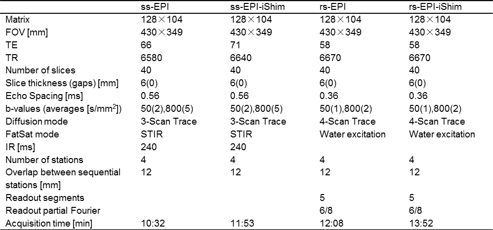
The iShim scheme was used to generate a non-product version of a commercial rs-EPI sequence (RESOLVE, Siemens Healthcare), referred as rs-EPI-iShim. The study was performed on a commercial 3 Tesla scanner (MAGNETOM Spectra, Siemens Healthcare, Erlangen, Germany) equipped with a 20-channel head, a 24-channel spine coil and three 6-channel body coils. Axial DW images from a healthy volunteer were acquired with ss-EPI, rs-EPI, ss-EPI-iShim (non-product version) and rs-EPI-iShim protocols. Imaging parameters for in-vivo scans were shown in Table 1. It notes that different fat suppression techniques used in ss-EPI and rs-EPI: fat suppression with short TI inversion recovery (STIR) for ss-EPI as a routine way, water-selective excitation (WE) for rs-EPI to compensate for the inherent longer acquisition time of the multi-shot acquisition in rs-EPI.RESULTS
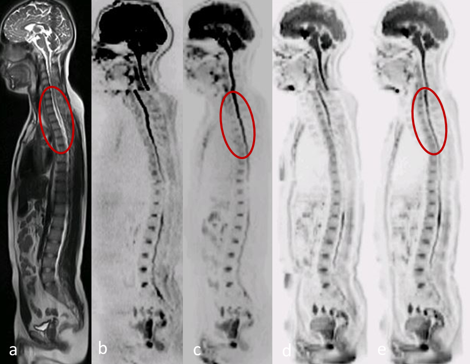
Fig.1 compares the reformatted sagittal MPR sample images (b = 800 s/mm2) acquired using ss-EPI, ss-EPI-iShim, rs-EPI and rs-EPI-iShim. As expected, the “broken spine” artifact was obviously visible in both ss-EPI (Fig.1b) and rs-EPI (Fig.1d), worse in ss-EPI. After the application of iShim, both ss-EPI-iShim (Fig.1c) and rs-EPI-iShim (Fig.1e) showed a remarkable reduction in distortions and signal loss. Considering the distortions, rs-EPI-iShim showed the best overall image quality. For example, compared with a T2 weighted HASTE reference image (Fig.1a), the spine region indicated with red circles is much less distorted with rs-EPI-iShim than with ss-EPI-iShim. However, the discontinuity could not be totally eliminated in both ss-EPI-iShim and rs-EPI-iShim. This might be due to the distortions introduced by high order component of the static field inhomogeneity.DISCUSSION
Spectrally attenuated inversion recovery (SPAIR) and chemical shift-based fat suppression that is commonly used in clinical DWI acquisitions employ spatial non-selective pulses and are therefore not compatible with slice-specific shimming (e.g. iShim). WE and STIR are compatible with iShim. STIR is insensitive to the static field inhomogeneity and has the best fat suppression performance, but will lead to SNR drops compared with other fat suppression methods. In this study, WE with reduced number of averages was used for rs-EPI and rs-EPI-iShim to compensate for the longer acquisition time in rs-EPI. So the selection of fat suppression method will also have an effect on the performance of iShim based WB-DWI. In the study, the fat suppression in ss-EPI based scans outperforms the other ones but has a lower SNR compared with that of rs-EPI based scans. More investigations will be done to find the best way of the fat suppression method in WB-DWI combined with iShim.CONCLUSION
Conventional frequency adjustment
and 3D shimming is performed per station and used for the entire slice stack of
each position, which is not applicable for the regions with larger variation of
center frequency shift and linear frequency shift. With iShim scheme, center
frequencies and linear shim terms could be dynamically updated prior to the
acquisition for each slice which leads to a significant reduction of susceptibility
related artifacts in these complex regions 4. Compared with
ss-EPI-iShim, rs-EPI-iShim could provide higher quality WB-DWI, specifically less
spatial distortions.CONCLUSION
Acknowledgements
No acknowledgement found.References
1. Padhani AR. et al. Whole-body diffusion-weighted MR imaging in cancer: current status and research directions. Radiology. 2011; 261:700-718.
2. Koh DM. et al. Whole-Body Diffusion-Weighted MRI: Tips, Tricks, and Pitfalls. American Journal of Roentgenology. 2012; 199:252-262.
3. Stemmer A, Kiefer B. Combination of integrated slice-specific dynamic shimming and pixel-wise unwarping of residual EPI distortions. Proc Intl Soc Mag Reson Med. 2015; 23:3729.
4. Zhang H. et al. Integrated shimming improves lesion detection in whole-body diffusion-weighted examinations of patients with plasma disorder at 3 T. Invest Radiol. 2015; 51(5): 297-305.
5. Porter DA, Heidemann RM. High resolution diffusion-weighted imaging using readout-segmented echo-planar imaging, parallel imaging and a two-dimensional navigator-based reacquisition. Magn Reson Med. 2009; 62(2):468–75.
6. Sven S. et al. Combination of integrated dynamic shimming and readout-segmented echo planar imaging for diffusion weighted MRI of the head and neck region at 3 Tesla. Magn Reson Imaging. 2017; 42:32-36.
Figures