3191
Improved brain MR imaging from a compact, lightweight 3T scanner with high performance gradients.1Mayo Clinic, Rochester, MN, United States, 2GE Global Research, Niskayuna, NY, United States
Synopsis
A compact, low-cryogen 3T MRI scanner with high-performance gradients capable of simultaneously achieving 80 mT/m and 700 T/m/s was compared to a 60-cm, whole body 3T system (50 mT/m, 200T/m/s) for 5 routine brain MR imaging sequences in 9 clinical patients, graded by two neuroradiologists. The compact 3T system performed equally well to a standard whole-body scanner in terms of motion artifacts, and performed better in terms of signal-to-noise ratio, lesion conspicuity, gray/white contrast, susceptibility artifacts and overall exam quality.
Purpose
The purpose of this NIH-funded initiative was to design, build, and evaluate a high-performance, low-cryogen, compact 3T (C3T) MRI system with high-performance gradients, capable of scanning heads, extremities and infants1,2,3. The reduced size, low liquid helium (<12 L) and light weight of the compact 3T system offers ease of siting. The initial goal was to show parity with a whole body system in terms of image quality and acquisition time. We previously reported findings of superior imaging with the compact 3T compared to a whole body system when evaluating a sagittal T2-weighted 3D FLAIR sequence in 16 human volunteers4. This work now extends the evaluation to other brain MRI sequences (T1 MP-RAGE, 3D T2 FLAIR, T2 FSE, GRE EPI, and DWI) obtained in 9 previously-unreported, patient subjects undergoing routine clinical imaging of the brain.Methods
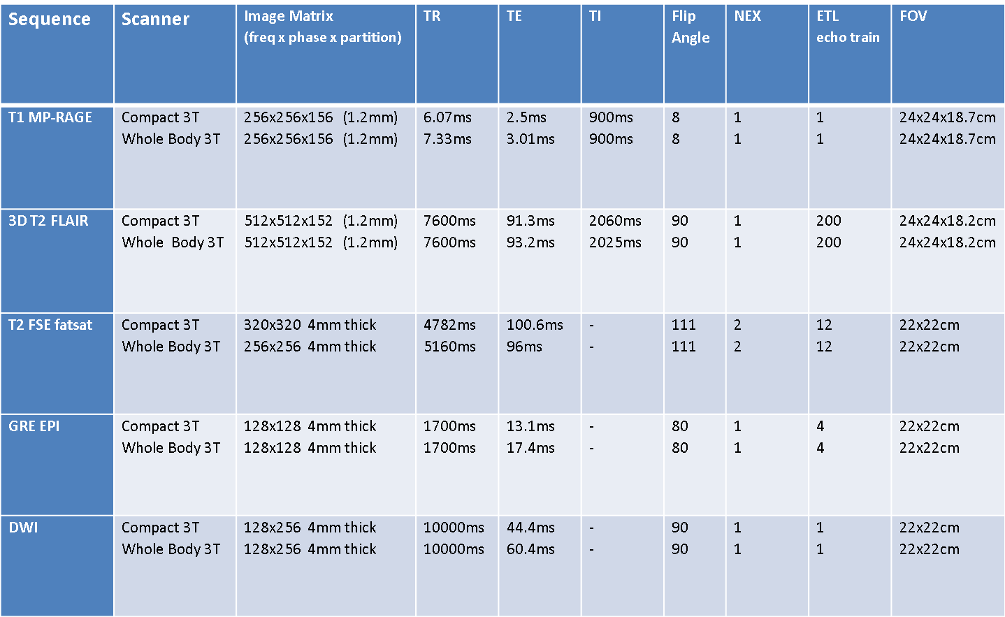
Under an IRB-approved protocol, 9 clinical patients undergoing a head MR examination consented to be scanned both on a standard, 60-cm bore, whole-body MR system with 50 mT/m, 200 T/m/s gradients (GE Discovery MR750, GE Healthcare, Waukesha, WI) and the compact 3T (C3T) with 80 mT/m 700 T/m/s gradients using an 8-channel receiver coil (In-vivo, Gainesville, FL). Imaging sequences evaluated included sagittal T1-weighted 3D MP-RAGE, sagittal T2-weighted 3D FLAIR, axial T2 FSE, axial GRE EPI, and DWI. Parameters used for these pulse sequences are summarized in Table 1. The C3T used real-time gradient pre-emphasis5 and frequency shifting to compensate additional concomitant fields arising from the asymmetric design of the transverse gradient coils. The 3D T2 FLAIR and T1 MP-RAGE sequences used a self-calibrating data-driven parallel imaging (ARC)6 acceleration factor of R = 2. The imaging studies were reviewed by two board-certified neuroradiologists with 19 and 27 years of experience. They were graded on a five-point ordinal scale from -2 to +2, with +2 indicating strong preference for the compact system and -2 indicating strong preference for the whole-body system, +1 and -1 representing preference for C3T or whole-body system respectively, and 0 representing no system preference. Each pair of images was comparatively evaluated (non-blinded) using the following attributes: signal-to-noise ratio (SNR), lesion conspicuity, motion artifact, gray/white matter contrast, susceptibility artifacts, cerebellar folia conspicuity, and overall exam quality. Statistical analysis was performed using a one-sided Wilcoxon signed rank tests7. The null hypothesis of the left-sided test was that the C3T performed equally to or better than the standard whole-body 3T MR system; and the right-sided test used the reverse of this hypothesis.Results
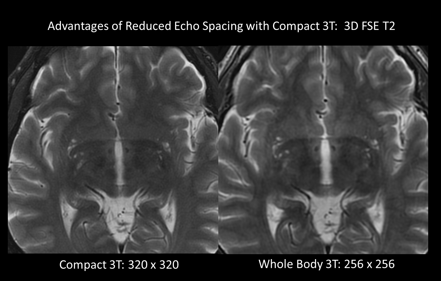
Figure 1 shows
comparison images from the two scanners obtained in the same patient with small
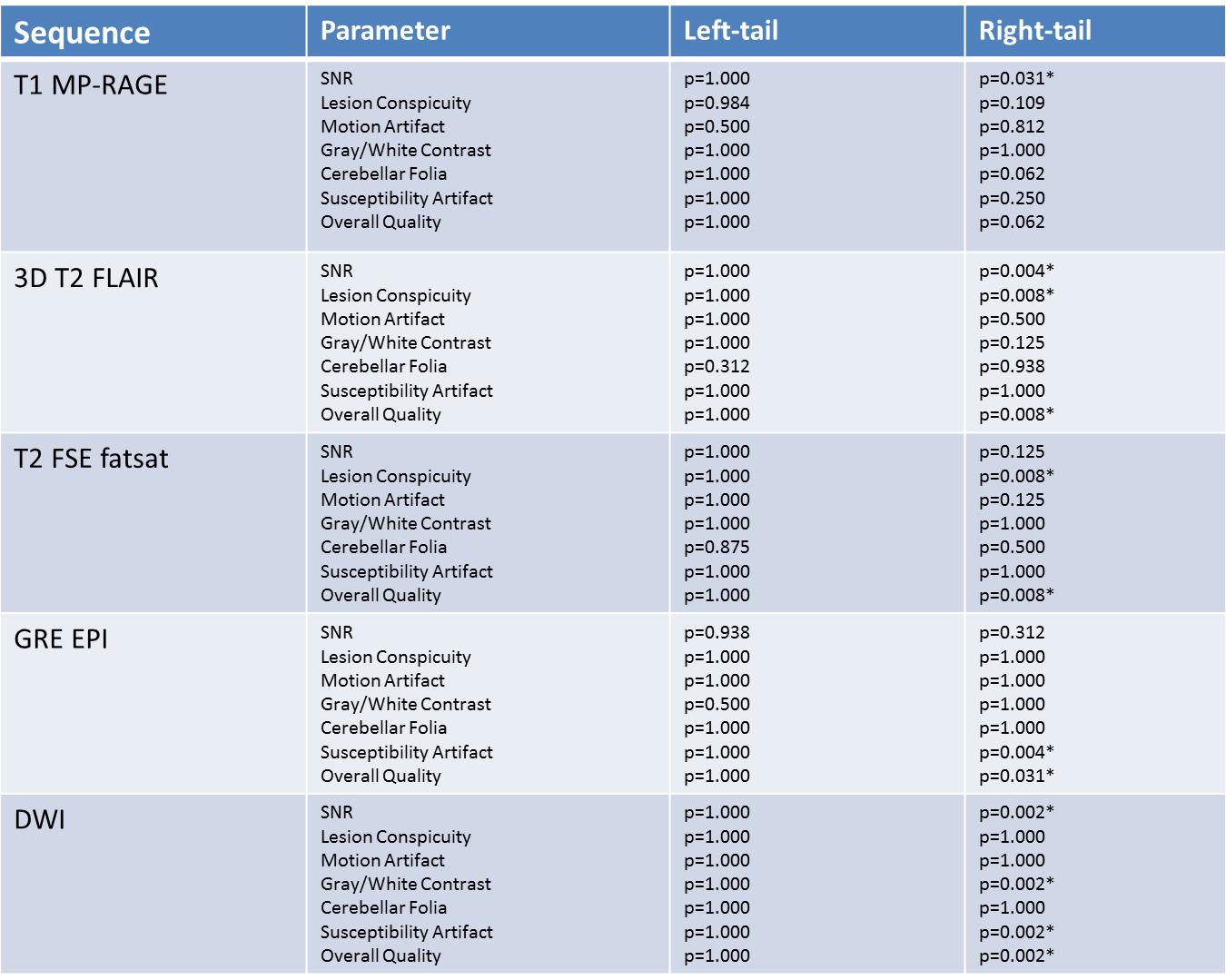
vessel ischemic disease. Table 2 shows summary of the results for left and
right tailed Wilcoxon signed-rank statistics for all evaluated parameters
across the five imaging sequences. Presuming a 5% significance level, the
right-sided test results indicate that the C3T outperformed the 60-cm bore whole
body system in terms of overall quality for all of the evaluated sequences,
except the T1-weighted MP-RAGE. All left-sided tests also failed to reject the
null hypothesis, indicating that the compact 3T system performed as good as the
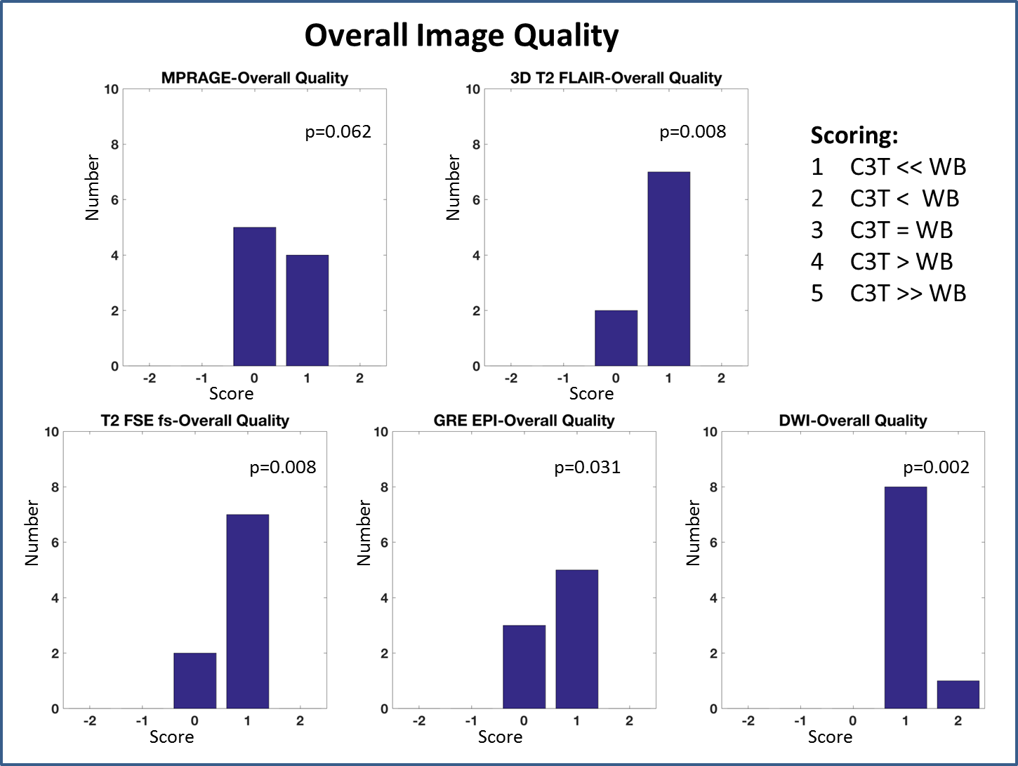
whole-body system for all five evaluated sequences. Histograms showing pooled
results for overall quality comparison between the C3T and body systems are
presented in Figure 2, showing comparison results for the five evaluated
imaging sequences. The significance results of the right-tailed test are
overlaid onto these histograms in Figure 2.
Discussion
There is strong statistical evidence that the C3T provides equal or better image quality for the five of the most commonly performed brain pulse sequences evaluated across this patient sample set. The unchanged incidence of motion artifact also suggests that the compact system offers at least equivalent patient comfort. The high slew-rate gradient system of the C3T scanner allows for an echo spacing reduction of 25% compared to the whole-body system. For a long echo train length sequence such as 3D T2 FSE, this echo space reduction can be used to acquire higher resolution images while maintaining similar readout duration and SNR (Figure 3).Conclusion
The compact, low-cryogen 3T MRI scanner with high-performance gradients provided equal or better image quality for five standard clinical brain MR imaging sequences in a small patient cohort.Acknowledgements
This work was supported in part by NIH grants BRP-R01-EB010065 and U01 EB024450.References
[1] Lee S-K, Mathieu J-B, Graziani D, Piel J, Budesheim E, Fiveland E, et al. Peripheral nerve stimulation characteristics of an asymmetric head-only gradient coil compatible with a high-channel-count receiver array. Magn Reson Med. 2016; 76:1939-1950.
[2] Mathieu J-B, Lee S-K, Graziani D, Lin J, Budesheim E, Piel JE, et al. Development of a Dedicated Asymmetric Head-only Gradient Coil for High-Performance Brain Imaging with a High PNS Threshold. Proceedings of the ISMRM Annual Meeting, Toronto: 2015, p. 1019.
[3] Weavers PT, Shu Y, Tao S, Huston J, Lee S-K, Graziani D, et al. Technical Note: Compact three-tesla magnetic resonance imager with high-performance gradients passes ACR image quality and acoustic noise tests. Med Phys 2016;43:1259–64. doi:10.1118/1.4941362.
[4] Weavers PT, Campeau NG, Shu Y, Tao S, Trzasko JD, Gray EM, Foo TK, Bernstein MA, Huston J. Improved T2-weighted 3D FLAIR from a compact, lightweight 3T scanner with high performance gradients. Proceedings of the ISMRM Annual Meeting, Honolulu, Hawaii:2017.
[5] Tao S, Weavers PT, Trzasko JD, Shu Y, Huston J, Lee S, et al. Gradient pre-emphasis to counteract first-order concomitant fields on asymmetric MRI gradient systems. Magn Reson Med. 2017, 77:2250-2262.
[6] Brau ACS, Beatty PJ, Skare S, Bammer R. Comparison of reconstruction accuracy and efficiency among autocalibrating data-driven parallel imaging methods. Magn Reson Med 2008;59:382–95.
[7] Siegel S. Nonparametric Statistics for the Behavioral Sciences. McGraw-Hill; 1956.
Figures