3180
Utility of real-time field control in subjects with mild cognitive impairment: T2* weighted imaging at 7T.1ETH & University Zurich, Zurich, Switzerland, 2University of Zurich, Zurich, Switzerland, 3University of Bern, Bern, Switzerland
Synopsis
Subjects with Alzheimer disease or mild cognitive impairment are known to produce strong field perturbation by particular breathing pattern and limb motion. In this work we explore the feasibility and success of real-time field control in subjects with mild cognitive impairment and age-matched controls in the scenario of high resolution T2*-weighted imaging at 7T. Real-time field control shows to be feasible and yield greatly enhanced data quality in both type of subjects: mild cognitive impairment and cognitively normal.
Introduction
T2*-weighted imaging in subjects with Alzheimer disease (AD) or Mild Cognitive Impairment (MCI) is hindered by particularly strong physiologically-induced field perturbations (arm motion, swallowing, and breathing)1. Such perturbation are believed to be the principal source of the increased artefact level seen in these subjects1.
Recently, real-time field control2, a method based on real-time field sensing, was shown to compensate for field perturbation and improve structural T2*-weighted imaging quality in healthy subjects3. The feasibility of this method in challenging conditions such as scanning of MCI subjects remains an open question.
In this work, we look at the feasibility of real-time field control in the realistic clinical scenarios of T2*-weighted acquisitions in MCI and Cognitively Normal (CN) elderly controls.
Method
The participants received medical, neurological and psychiatric examination, as well as neuropsychological testing during screening for eligibility to participate in the study. They were categorized as either cognitively normal (CN) or mild cognitive impairment (MCI) according to established criteria for diagnosis of MCI4,5. The study group consisted of 39 participants, aged between 50 and 96 years old (29CN/10MCI).
A high resolution 3D T2*-weighted structural scan (resolution:0.6x0.6x2mm3; FOV:200x180.72x130.2mm3; TR/TE/flip angle:40ms/14ms/10°; duration:12min34sec) was acquired on a 7tesla (Philips, Achieva) and reconstructed on the scanner consol. The scanner was equipped with a full third-order shimming system.
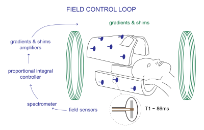
Field sensing was performed with long-lived NMR field probes6 arranged in a cylindrical manner2. Field values were acquired for every other slice TR, 7ms prior to the next slice excitation and after the readout. Each measurement was streamed to a proportional-integral controller, which computed the shim input update necessary to counter the measured perturbation. The shim channels were consequently actuated (figure 1).
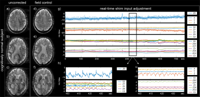
The measured field deviation traces were inspected to confirm the correction efficacy. The applied correction consists of 16 shim input traces (0th to 3rd order). These traces were scaled to the maximum field offset produced by each coils over a virtual 20cm diameter sphere (Hz Max).
Results
Uncorrected and corrected data of a CN subject are presented in figure 2. Uncorrected data (Fig2a-c) are affected by strong shading artefacts. Corrected data (Fig2f-h), on the other side, do not exhibit shading artefacts yielding high data quality. Correction time courses for the whole acquisition (Fig2g) exhibit short term (modulation) and long term (drift) variations. Taking a closer look, breathing and heart beat related field changes become visible (Fig2h). For example, in the ZX trace (Fig2h), slow modulation relate to breathing and faster modulation relate to the heart beating. On the other side, only heart beat seem to affect the S2 trace.
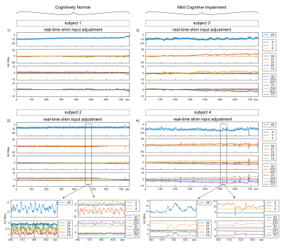
Shim actuation time courses for CN and MCI subjects are presented in figure 3. While cognitively normal subjects breath regularly, MCI subjects tend to have an irregular breathing cycle and a larger breathing amplitude.
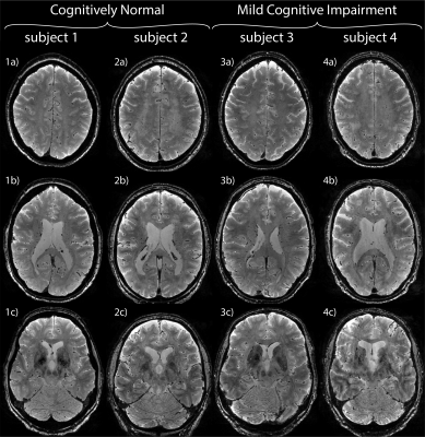
The resulting T2*-weighted data are shown in figure 4. In all four subjects, the correction was successful. In the lowest slice of subject 4, light shading indicate the presence field perturbations of higher order than the three orders covered by the field control.
Discussion
Field control was shown to be equally feasible in MCI and cognitively normal subjects. Physiology related perturbation were successfully mitigated. This success was reflected in the enhanced imaging results. The correction time courses showed to be rich in physiological information such as breathing and heart beat. Generally, MCI correction time courses are more complex in their dynamics than time courses of CN subjects. The system being limited to third order spherical harmonics, any field perturbations of orders higher than three cannot be effectively addressed. Obviously, head motion remains a challenge as MCI and AD subjects encounter increased difficulties to cooperate over prolonged scan time. A method combining field control and motion correction was demonstrated in healthy controls and remain to be tested in the challenging scenario of MCI and AD7.Conclusion
By demonstrating that field control is feasible and yield enhanced image quality in a clinical study setting, this approach holds promises for clinical application. We observe a prevalence of irregular field perturbations for MCI subjects as in Versluis et al.1. It confirms that MCI subjects have difficulties lying still (limb motion).Acknowledgements
No acknowledgement found.References
1. Versluis et al. Origin and reduction of motion and f0 artifacts in high resolution T2*-weighted magnetic resonance imaging: Application in Alzheimer’s disease patients. Neuroimage 51 (2010)
2. Duerst et al. Real-Time Feedback for Spatiotemporal Field Stabilization in MR Systems. MRM 73 (2015)
3. Duerst et al. Utility of Real-Time Field Control in T2*-Weighted Head MRI at 7T. MRM 76 (2016) 4. Petersen et al. Mild Cognitive Impairment: clinical characterization and outcome. Arch Neurol 56 (1999)
5. McKahn et al. The diagnosis of dementia due to Alzheimer’s disease: Recommandations from the National Instituted on Aging-Alzheimer’s Association workgroups on diagnostic guidelines for Alzheimer ‘s disease. Alzheimer’s Dement 7 (2011)
6. De Zanche et al. NMR probes for measuring magnetic fields and field dynamics in MR systems. MRM 60 (2008)
7. Vionnet et al. Simultaneous prospective motion correction and feedback field control: T2* weighted imaging at high field, 306, 25th ISMRM, 2017.
Figures


