3176
Imaging Ultrashort-T2* structures in the Eye with UTE MRI1Radiology and Biomedical Imaging, University of California - San Francisco, San Francisco, CA, United States, 2Ophthalmology and Radiology, New York University, New York, NY, United States
Synopsis
Ultrashort echo time (UTE) MRI has the potential to image all structures in the eye, including the sclera, cornea and lens that have relatively short T2* relaxation times. UTE MRI was used for motion-robust imaging of the eye in vivo, and multiple TE measurements were combined for 3D T2* mapping. We report measurements of in vivo T2* relaxation times in the range of several ms that suggest that UTE MRI can be used effectively to study ocular structures with short T2* in vivo.
Introduction
The microstructural organization and composition of the corneoscleral shell determine the biomechanical behavior of the eye, and are important in diseases such as glaucoma and myopia. The sclera and cornea in the corneoscleral shell are dense and fibrous connective tissues that form the outer coat of the eye, which act to support and protect the eye from the surrounding environments. Apart from the corneoscleral shell, the lens in the eye consists of tightly packed fibers, and lens opacifications during aging and cataract formation are often accompanied by alterations in tissue compositions including the protein hydration state. These structures are difficult to image with conventional MRI, both due to motion as well as short relaxation times in protein-rich tissues [1].
Ultrashort echo time (UTE) MRI has the potential to image all structures in the eye, including the sclera, cornea and lens that have relatively short T2* relaxation times. Eye imaging in vivo is also challenging due to motion such as blinking and eye movements during gaze that can easily lead to motion artifacts. UTE MRI is advantageous as well since it is inherently robust to motion [2], thus it is expected to be less corrupted by eye motion. In this study, we examine the feasibility of in vivo 3D UTE MRI relaxometry in healthy adult human eyes.
Methods
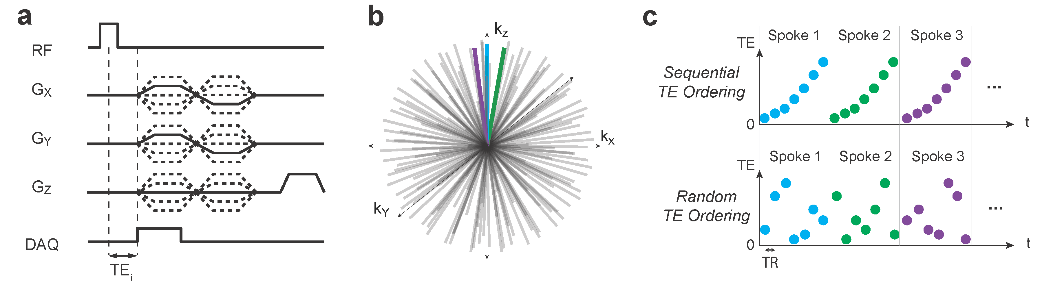
Ultrashort-T2* relaxometry was performed using a 3D UTE pulse sequence with the delay between excitation and readout shifted between TRs to acquire a set of 6 or 7 TEs within a single scan (Figure 1). All TEs were ≤ 5 ms to characterize the ultrashort-T2 components, with a relatively larger proportion of TEs under 1 ms. Scan times were ~20 minutes for 1mm isotropic resolution at TR = 10ms, and ~10 minutes for 2mm isotropic resolution at TR = 6ms. Data was acquired on a 3T GE MRI systems, with a 32-channel head coil (Nova Medical). A non-Cartesian ESPIRiT parallel imaging reconstruction method [3] was implemented using BART (https://mrirecon.github.io/bart/) with acceleration factors from R=4 to 6. A randomized TE ordering was used to remove eddy current artifacts.
T2* was estimated by fitting to an exponential model that also included a term to account for off-resonance. This was applied to the complex valued images.
Results
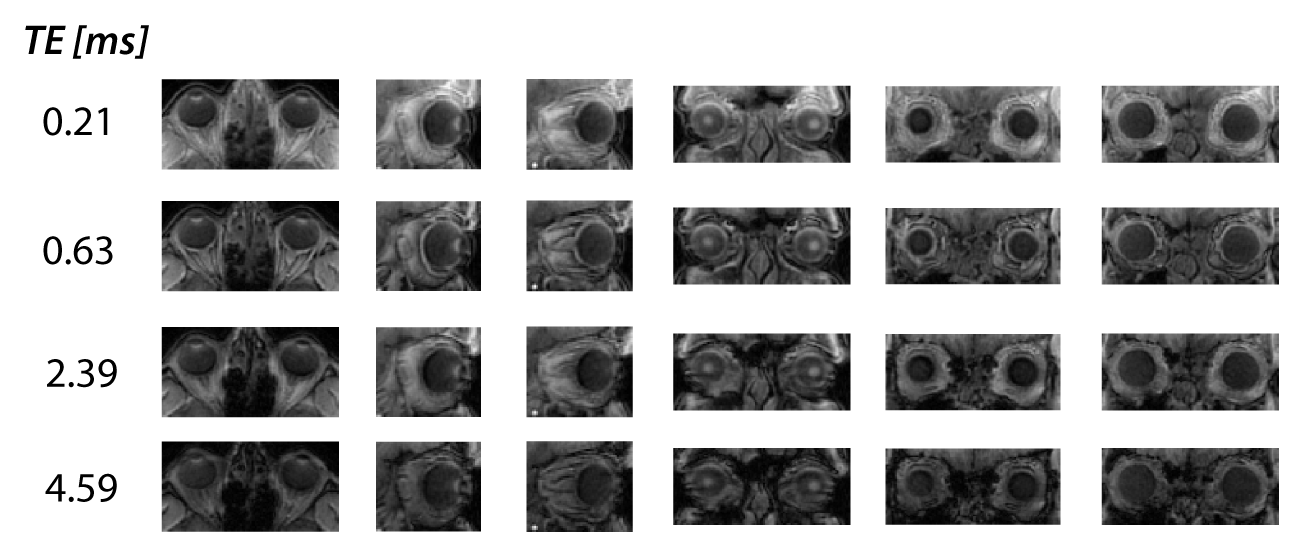
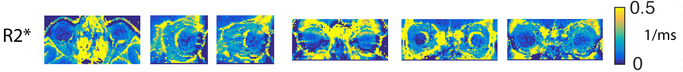
Images at various TEs show some clear signals around the eye that decays by the later echo times (Figure 2). This structure, which appears to correspond to the corneoscleral shell, was measured to have a T2* as short as 1.4-1.8 ms in the posterior portion of the eye, although elsewhere was observed to have a longer T2* up to 5 ms (Figure 3). At TE = 0.6 ms there are some chemical shift artifacts due to out of phase fat and water signals.
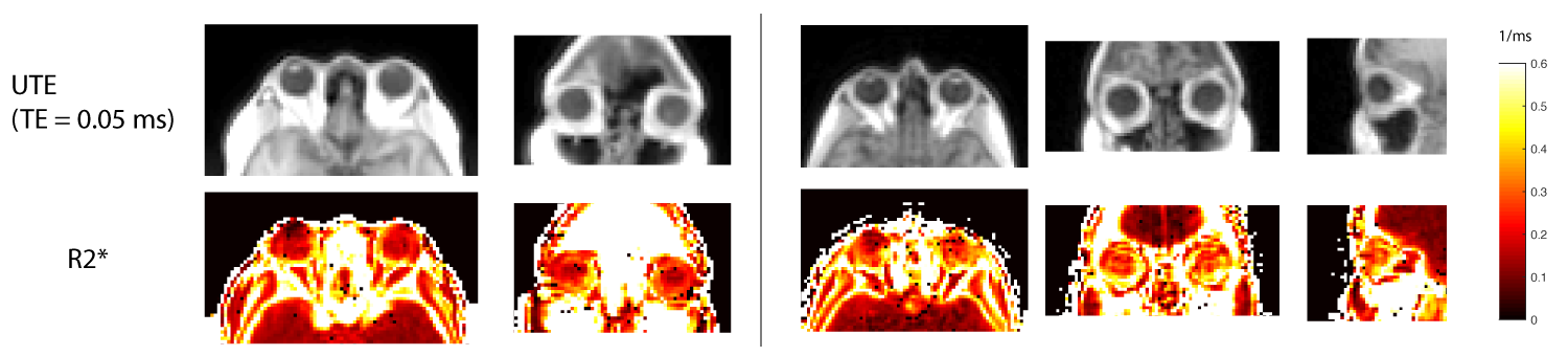
From the 2mm isotropic, whole-head scan (Figure 4), T2* relaxation times were measured to be > 5 ms within the vitreous body. Around the vitreous body T2* was measured to be approximately 2-3 ms, which likely includes the sclera and retina surrounding the eye. We also observed similar T2* values at around 2 ms in the optic nerve.
Conclusion
UTE MRI was used for motion-robust imaging of the eye in vivo, and multiple TE measurements were combined for 3D T2* mapping. We report measurements of in vivo T2* relaxation times in the range of several ms that suggest that UTE MRI can be used effectively to study ocular structures with short T2* in vivo.Acknowledgements
No acknowledgement found.References
1. Ho LC, Sigal IA, Jan N-J, Yang X, Merwe Y van der, Yu Y, Chau Y, Leung CK, Conner IP, Jin T, Wu EX, Kim S-G, Wollstein G, Schuman JS, Chan KC. Non-invasive MRI Assessments of Tissue Microstructures and Macromolecules in the Eye upon Biomechanical or Biochemical Modulation. Sci Rep. 2016 Aug 26;6:srep32080.
2. Jiang W, Ong F, Johnson KM, Nagle SK, Hope TA, Lustig M, Larson PEZ. Motion robust high resolution 3D free-breathing pulmonary MRI using dynamic 3D image self-navigator. Magn Reson Med. 2017.
3. Uecker M, Lai P, Murphy MJ, Virtue P, Elad M, Pauly JM, Vasanawala SS, Lustig M. ESPIRiT–an eigenvalue approach to autocalibrating parallel MRI: where SENSE meets GRAPPA. Magn Reson Med. 2014 Mar;71(3):990–1001. PMID: 23649942
Figures