3164
Phase Laplacian Coil Combination1MR Physics, German Center for Neurodegenerative Diseases (DZNE), Bonn, Germany, 2Department of Physics and Astronomy, University of Bonn, Bonn, Germany
Synopsis
We propose a novel, computationally efficient coil combination technique for multi-channel phase data based on the Laplacian of single-channel phase images. This renders explicit knowledge or estimation of the receive sensitivities unnecessary. The combined phase Laplacian can be either be transformed back to unwrapped phase domain (Laplacian-based unwrapping) or directly utilized for further analyses based on the phase Laplacian, e.g. harmonic background-field removal. At 3T we demonstrate similar-to-improved phase reconstruction compared to the vendor-provided state-of-the-art coil combination, which uses the body-coil as a uniform reference, and successfully apply the technique at 7T.
INTRODUCTION
To obtain an accurate phase representation of MR images, one optimally combines phased array data by a weighted sum of all complex single-coil images, essentially using the complex conjugate receive sensitivity (RX) profiles as weights1. In the absence of a homogenous “body coil”, typically used to estimate the RX profiles, virtual reference coil methods tend to fail at ultra-high fields (e.g. open ended fringe lines)2. Many state-of-the-art phase analysis techniques, however, require only the Laplacian of the phase field (LAP), e.g. (V)SHARP3,4, HARPERELLA5, LBV6, Laplacian-based phase unwrapping (LU)7,8 or improved preconditioned conjugate gradient Laplacian unwrapping (pccgLU)2. We propose to perform multi-channel phase combination efficiently in the LAP domain, rendering knowledge of receive sensitivities unnecessary.METHODS
Let $$$\phi_c = \alpha + \beta + \gamma_c + \eta_c$$$ be the phase image acquired by coil $$$c$$$ of the phased array. The first two terms are the internal, $$$\alpha$$$, and the background phase, $$$\beta$$$ (incl. RF transmission phase9). Coil-specific contributions are represented by the RX phase, $$$\gamma_c$$$, and phase noise, $$$\eta_c\in(-\pi,\pi]$$$. We assume that $$$\gamma_c$$$ is largely harmonic, i.e. any non-harmonic contributions are negligible or cancel partially by a weighted sum across all single-coil phase Laplacians:
(1) $$$\quad \Delta\Phi = \sum_c w_c\Delta\phi_c = \Delta(\alpha+\beta) + \sum_c w_c \Delta\eta_c + \sum_c w_c \Delta\gamma_c$$$
To minimize total noise, we propose to employ normalized weights according to squared coil magnitudes (Gaussian-smoothed, 1 voxel kernel width): $$$w_c = m_c^2/\sum_c m_c^2$$$. The last term in Eq. (1) can be regarded as a residual (non-harmonic) RX term.
Experiment:
Five healthy, young subjects underwent two consecutive whole-brain 3D-GRE measurements on a Siemens MAGNETOM Skyra 3T scanner. The latest software version (VE11C) provided state-of-the-art complex coil combination (32 channel head coil) utilizing the body coil as a reference. Imaging parameters were: 232x256x160 matrix, 0.9mm iso, GRAPPA R=2x1, TE=20ms, TR=27ms. Additionally, one subject was scanned in a Siemens MAGNETOM 7T research scanner using a comparable 32 channel coil, but lacking a body coil. A motion-robust dual-echo 3D-GRE-EPI sequence was employed10 (240x240x160 matrix, 0.8mm iso, GRAPPA R=3x1, Partial Fourier 7/8x1, TE1=11ms, TRvol=45s, 9 averages).
Analysis:
The Laplace operation was implemented as $$$\Delta\phi = \text{div}(b\ \text{grad}(\phi))$$$, where $$$b$$$ is a brain binary mask2 and $$$\phi$$$ can be the wrapped scanner-combined phase or a wrapped single-channel phase. For 3T(7T) data, $$$b$$$ was obtained by eroding a BET11 mask (from the root-sum-of-squares magnitude image) by 2(6) voxels. At this stage, $$$b$$$ is required only for pccgLU (here with 5 iterations). For simplified background-field removal (SHARP)4, the BET mask was eroded by 4(12) voxels and applied to LAP before inversion (non-regularized Laplace inversion based on discrete cosine transforms7).
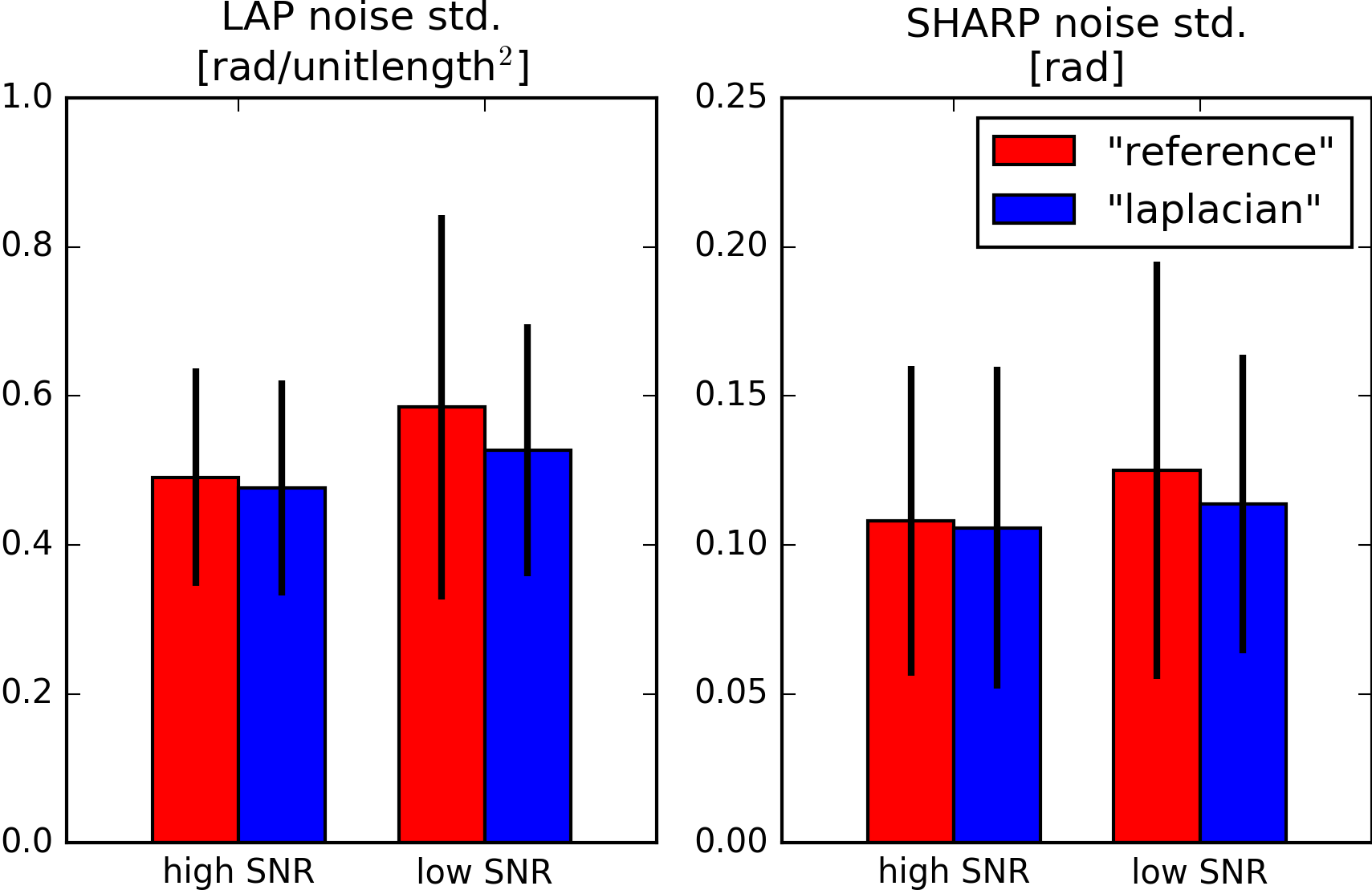
For the vendor-provided (“reference”) and the proposed coil combination (“laplacian”) the standard deviation of the LAP and SHARP noise distribution was evaluated in two regions-of-interest (ROI) from the difference12 between two separate LAP and SHARP maps. One peripheral brain ROI (“high SNR”, right occipital lobe) and one central brain ROI (“low SNR”) were defined on comparable, homogeneous white matter regions in all five subjects (5.4mm spherical ROI radius).
RESULTS & DISCUSSION
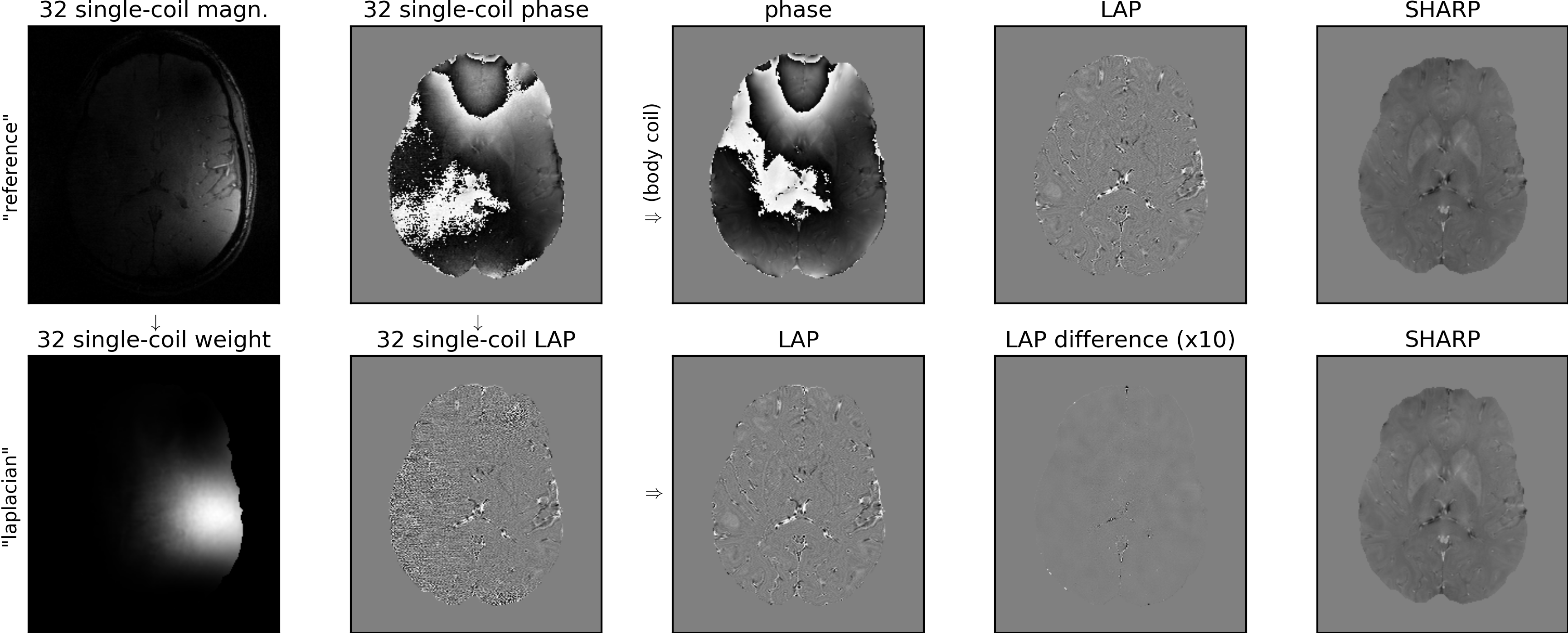
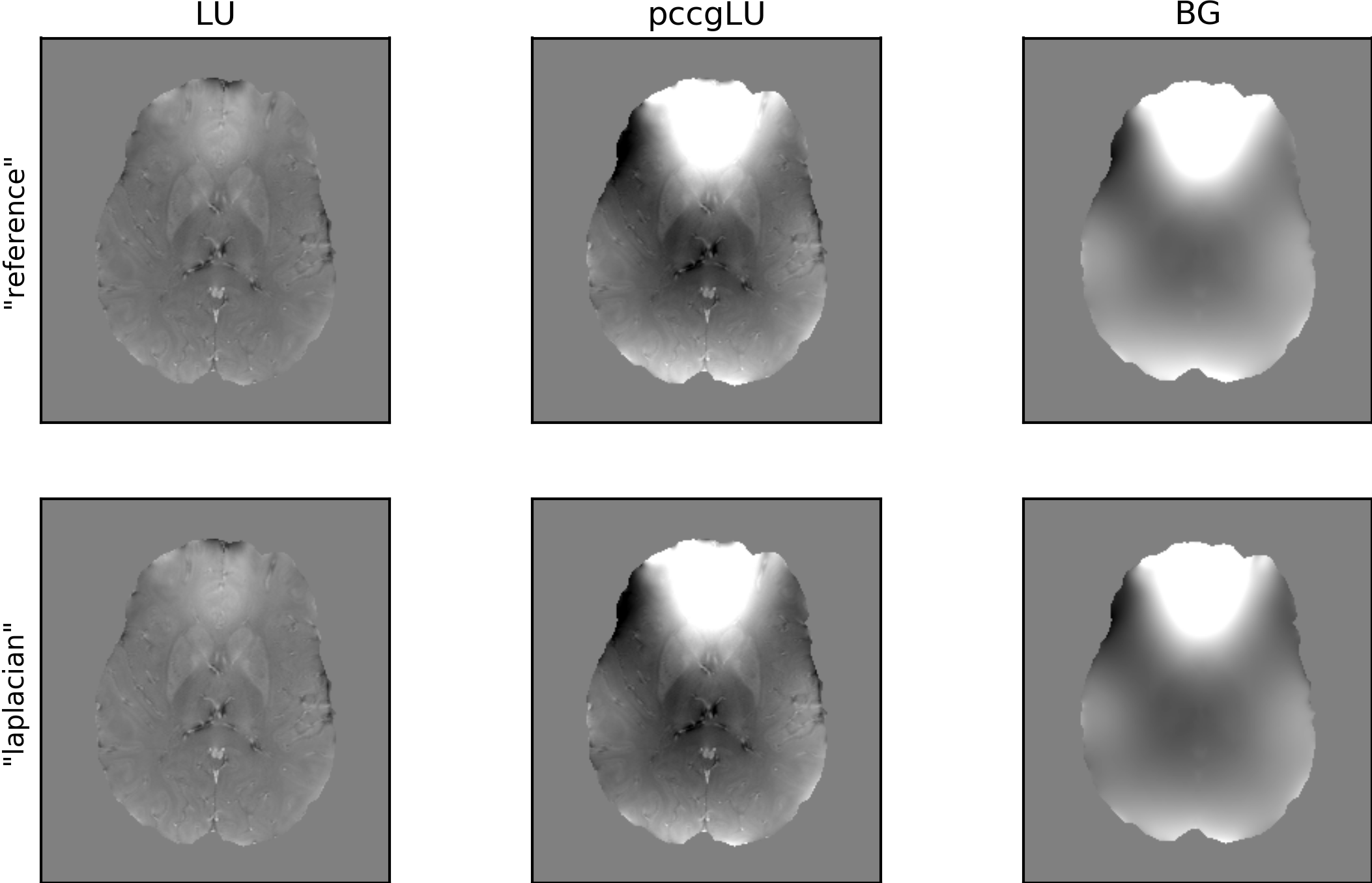
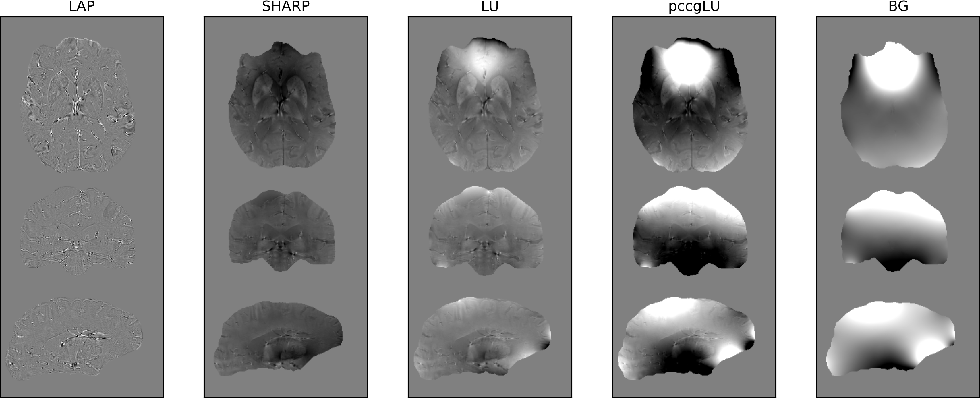
Fig. 1 illustrates, on one example subject and slice at 3T, the “reference” and the “laplacian” method and shows resultant LAP and SHARP maps. According to Eq. (1), the LAP difference map reveals noise and residual, non-harmonic RX differences between the “reference” and “laplacian” LAP maps (note different windowing). Group statistics at 3T, summarized in Fig. 2, indicate comparable noise propagation using either method. The “laplacian” method even seems to result in reduced noise, although the group size does not allow for statements on significance. The corresponding example (pccg)LU maps and the background field (“BG=pccgLU-SHARP”), displayed in Fig. 3, show very high agreement as well. Fig. 4 demonstrates that the proposed method can readily be applied at at ultra-high fields.CONCLUSION
We have proposed a novel multi-channel phase combination technique based on phase Laplacian images rather than on raw phase images. We have demonstrated similar or improved phase reconstruction (with or without background-field) compared to reference-based complex coil combination at 3T. The proposed method does not require a real or virtual reference coil. We could thus successfully apply it to 7T data. Future investigations will increase the group size and include virtual body coil methods in our comparison at 3T and 7T. Although only simplified background-field removal has been demonstrated here, the proposed method is also perfectly suited for advanced methods such as V-SHARP3 or for solving the Laplacian boundary value problem (LBV)6.Acknowledgements
No acknowledgement found.References
1 Roemer PB, Edelstein WA, Hayes CE. The NMR Phased Array. Magn Reson Med 1990;16(2):192-225.
2 Robinson SD, Bredies K, Khabipova D, Dymerska B, Marques JP, Schweser F. An illustrated comparison of processing methods for MR phase imaging and QSM: combining array coil signals and phase unwrapping. NMR Biomed 2016;(epub)(November 2015). https://doi.org/10.1002/nbm.3601.
3 Li W, Wu B, Liu C. Quantitative susceptibility mapping of human brain reflects spatial variation in tissue composition. Neuroimage 2011;55(4):1645-56. https://doi.org/10.1016/j.neuroimage.2010.11.088.
4 Schweser F, Deistung A, Sommer K, Reichenbach JR. Toward online reconstruction of quantitative susceptibility maps: Superfast dipole inversion. Magn Reson Med 2013;69(6):1582-94. https://doi.org/10.1002/mrm.24405.
5 Li W, Avram AV, Wu B, Xiao X, Liu C. Integrated Laplacian-based phase unwrapping and background phase removal for quantitative susceptibility mapping. NMR Biomed 2014;27(2):219-27. https://doi.org/10.1002/nbm.3056.
6 Zhou D, Liu T, Spincemaille P, Wang Y. Background field removal by solving the Laplacian boundary value problem. NMR Biomed 2014;27(3):312-9. https://doi.org/10.1002/nbm.3064.
7 Schofield M a, Zhu Y. Fast phase unwrapping algorithm for interferometric applications. Opt Lett 2003;28(14):1194-6. https://doi.org/10.1364/OL.28.001194.
8 Volkov VV, Zhu Y. Deterministic phase unwrapping in the presence of noise. Opt Lett 2003;28(22):2156-8. https://doi.org/10.1364/ol.28.002156.
9 Schweser F, Atterbury M, Deistung a, Lehr BW, Sommer K, Reichenbach JR. Harmonic phase subtraction methods are prone to B 1 background components. Proc 19th Sci Meet Int Soc Magn Reson Med 2011;37(9):6635.
10 Stirnberg R, Acosta-Cabronero J, Poser BA, Stöcker T.
2D-segmented, multi-TE 3D-EPI for high-resolution R2* and quantitative
susceptibility mapping at 7 Tesla. In
Proc Intl Soc Mag Reson Med 23, 2015.
11 Smith SM. Fast robust automated brain extraction. Hum Brain Mapp 2002;17(3):143-55. https://doi.org/10.1002/hbm.10062.
12 Dietrich O, Raya JG, Reeder SB, Ingrisch M, Reiser MF, Schoenberg SO. Influence of multichannel combination, parallel imaging and other reconstruction techniques on MRI noise characteristics. Magn Reson Imaging 2008;26(6):754-62. https://doi.org/10.1016/j.mri.2008.02.001.
Figures