3150
On Resonance Variable Delay Multiple Pulse (onVDMP) CEST MRI for Monitoring Stem Cell Therapy in Experimental Autoimmune Encephalomyelitis1Radiology, Johns Hopkins School of Medicine, Baltimore, MD, United States, 2Radiology, Kennedy Krieger Institute, Baltimore, MD, United States, 3Neurology, Johns Hopkins School of Medicine, Baltimore, MD, United States
Synopsis
In mice with experimental autoimmune encephalomyelitis, spatiotemporal signal changes were detected with on resonance variable delay multiple pulse (onVDMP) CEST MRI for the brain and spinal cord. Transplantation of glial-restricted precursor cells resulted in normalization of those signal changes during the pre-onset stage of the disease.
Purpose
To assess whether the onVDMP CEST MRI signal changes with the disease course of experimental autoimmune encephalomyelitis (EAE), an animal model of multiple sclerosis, and to evaluate its potential to monitor neurorestoration following stem cell transplantationMethods
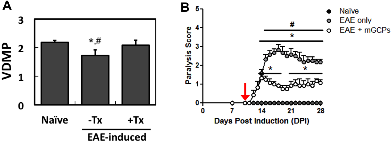
Induction of EAE: EAE was induced in C57Bl/6 mice (n=6-8) by i.p. injection of 250 ng of pertussis toxin and s.c. injection of 300 μg of MOG35-55 in IFA supplemented with 4 mg/ml tuberculin on day 0 and day 2. Mice were monitored daily and assessed for paralysis using the following clinical scoring system: 0=healthy, 1=tail paralysis, 2=mild hindlimb paralysis, 3=total hindlimb paralysis, 4=forelimb paralysis, and 5=moribund/death. Naïve mice were used as control (n=4-5).
onVDMP CEST MRI: Mice were imaged weekly using a horizontal bore Biospec 11.7T scanner. For transmission, a 72-mm volume resonator was used. For acquisition, 2x2 and 4x1 phased array coils were used for the brain and spinal cord, respectively. The slice thickness was 2 mm for all images. We used a onVDMP CEST MRI sequence that distinguishes fast exchanging protons1, with TE=5 ms, TR=5 s, Rare factor=10, NA=1, 9 repetitions, B1 = 46.8 μT, 32 pulse-exchange modules with a 2 ms pulse width, eight delays (mixing times from 1.14 to 100 ms), and FOV=128x128.
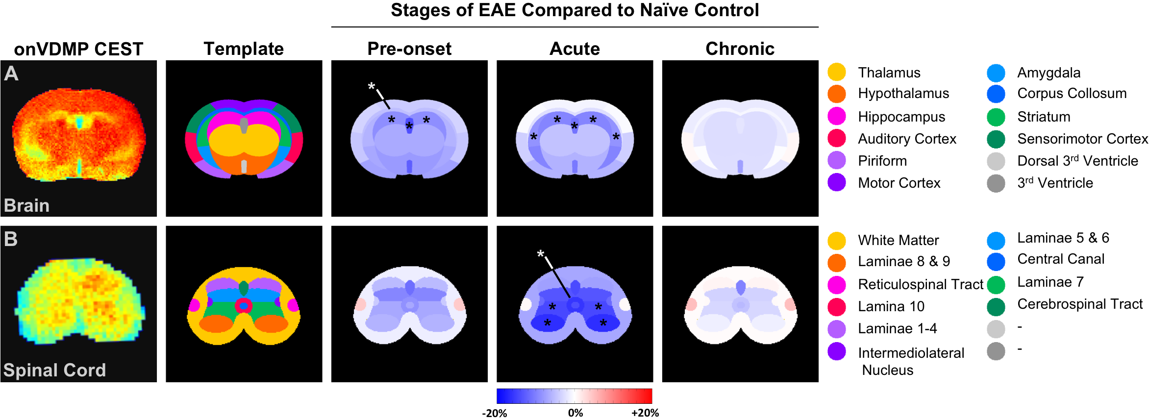
Variation map: Maps assessing regional changes in onVDMP CEST
signal during EAE compared to naïve controls were generated using a
segmentation template based on a mouse atlas (atlas.brain-map.org) and the
following formula:
100 * (onVDMPEAE STAGE – onVDMPNAÏVE)
/ onVDMPNAÏVE.
Cell culture: Murine glial-restricted progenitor cells (GPCs) were isolated at E14.5 from the CNS of luciferase- and PLP-eGFP transgene expressing neonatal pups and cultured for two weeks on PLL/laminin-coated plates in basal media supplemented with bFGF (20 ng/ml), N2 (2%), B27 (1%), and bovine serum albumin (0.1%).
Cell transplantation: Two million cells were injected intracerebroventricularly 6 days after EAE induction using a Hamilton 31G microinjection needle and a stereotaxic device.
Statistics: Significance was defined at p<0.05 using an ANOVA with Tukey post-hoc.
Results and Discussion
onVDMP CEST MRI revealed transient alterations in the ventricle, striatum, and laminae 8&9 of the ventral horn (Figure 1). Transplantation of GPCs into the lateral ventricle attenuated EAE based on onVDMP CEST signal and paralysis score (Figure 2).Acknowledgements
Funding was provided by the NMSS (RG 4994-A-3) and the TEDCO Maryland Stem Cell Fund for Postdoctoral Fellows.References
1. J Xu et al. Magn Reson Med. 2017;77(2):730-739.Figures