3139
Prediction of Alzheimer’s disease by using deep learning 3D-Convolutional Neural NetworksNa Sang1, Francisco M. Garcia2, Wanshun Wei3, Huabing Li4, Tao Ma1, and Silun Wang1
1YIWEI Medical Inc, Shenzhen, China, 2University of Massachusetts - Amherst, Amherst, MA, United States, 3YIWEI Meidcal Inc, Shenzhen, China, 4ZhongNan University, ChangSha, China
Synopsis
We analyzed the T1 structural MRI by using deep learning 3D-CNN method. The results indicate that deep learning models can accurately predict AD patients with diagnostic accuracy of 96%. This can be achieved using raw MRI data, with a minimum of processing necessary to generate an accurate AD prediction. Our model shows highly sensitivity and negative predictive value and thus appropriate for use for screening testing in population study. Currently model has the potential to be used as a screen biomarker to investigate the neurodegeneration, brain aging and associated brain diseases.
Introduction
Alzheimer’s disease (AD) is the most common form of dementia and its prevalence is set to rise in the coming decades1. It has been an incredible increase in performance in classification and regression models mainly sparked by deep learning techniques. One area in particular that has seen a dramatic improvement in performance is computer vision, through the use of convolutional neural networks (CNN) and its variants. We aim to predict the AD with a deep 3D convolutional neural network (3D-CNN), which can learn generic features capturing AD biomarkers and adapt to different domain datasets.Methods
Dataset: T1-weighted magnetic resonance imaging (MRI) data from 90 AD patients with mean age of 71.7 ± 5.9 years and 151 age and sex matched healthy controls (mean age = 71.4 years ±4.8) were obtained from the Alzheimer's Disease Neuroimaging Initiative (ADNI) database. Among them 80% AD patients and controls were recruited as training dataset and others were regarded as testing dataset. MRI data preprocessing: All MRI data were preprocessed by using the SPM8 software package (UCL, UK) which included tissue segmentation; registration and resampling. Each tissue class (i.e., GM and WM) was processed independently after segmentation (Figure1). Feature extraction and classification: Figure 2 and 3 show the architectures used for the convolutional autoencoder, and the full classifier neural network. Briefly, given an input image X with height h, width w, and depth d, let us call the dimensionality of X. We seek an enconder f(X) with dimensionality k, k << n, decoder g(f(X)), and train a model with weights W to minimize the objective , that is, the square-error between the decoding of the low dimensional encoding and the original input. Given this new representation f(X), we train a neural network classifier using f(X) as input.Results
Feature exaction: The autoencoder part of the network is able to generate low-dimensional representation of the input data, extracting in the process the features that is most useful to predicted the presence or absence of the disease. Figure 4 is an example of the generated 3D images from the autoencoder. Diagnostic accuracy: Our current system shows an average accuracy of 96% over our testing set doing 5-fold cross validation with sensitivity of 100%, specificity of 92%, positive predictive value of 90% and negative predictive value of 100%.Discussion & Conclusion
Deep learning models with 3D-CNN based on T1-MRI can accurately predict AD patients. This can be achieved using raw MRI data, with a minimum of processing necessary to generate an accurate AD prediction. These estimates of AD prediction model are also significantly heritable, giving external, genetic, validity to the measure and motivating its use in genetic studies of neurodegeneration diseases diagnosis. Finally, our model shows highly sensitivity and negative predictive value and thus appropriate for use for screening testing in population study. Currently model has the potential to be used as a screen biomarker to investigate the neurodegeneration, brain aging and associated brain diseases.Acknowledgements
No acknowledgement found.References
1. Alzheimer’s Association et al., “2014 alzheimer’s disease facts and figures,” Alzheimers Dement, vol. 10, no. 2, pp. e47–e92, 2014.Figures
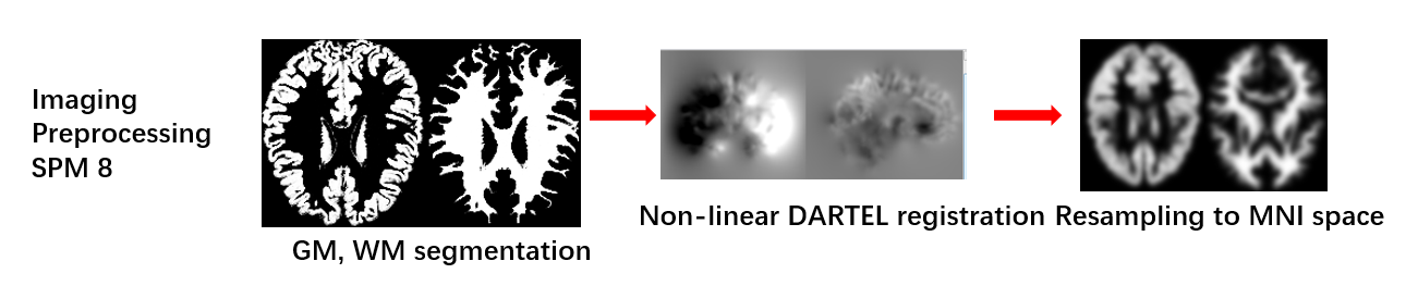
Imaging preprocessing
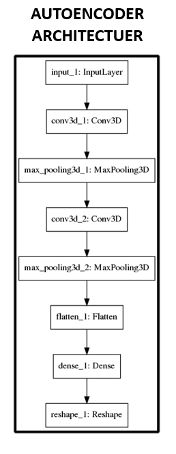
Architectures used for the convolutional
autoencoder
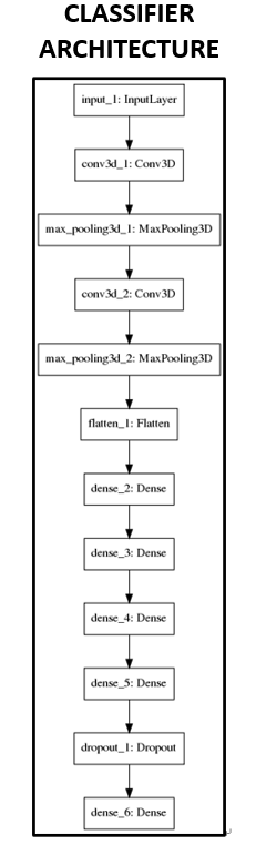
Architectures used for full classifier neural network
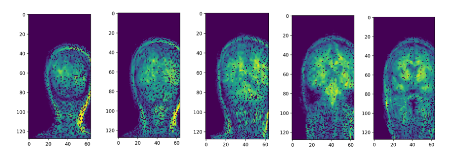
Figure 4 is an example of the generated 3D
images from the autoencoder