3136
Carotid Artery Segmentation Based on Level Set Method in MR Images1Department of Biomedical Engineering, Beijing Institute of Technology, Beijing, China, 2Center for Biomedical Imaging Research, Department of Biomedical Engineering, Tsinghua University School of Medicine, Beijing, China
Synopsis
Segmentation of blood vessels is the first step of visualization of vascular geometry and morphological analysis in cardiovascular diagnosis. However, the manual segmentation of blood vessels is time consuming. This study applied the level set method to automatic segmentation for carotid artery morphology on time-of-flight MR angiography images. The brightness weighting term is added to optimize the model, and the optimal setting of the parameters of the carotid artery images is discussed in detail. The results showed that the model with the brightness weighting term was more stable, accurate and efficient, which might be used to automatic segmentation of carotid arteries.
Introduction
It has been shown that geometric characteristics of carotid arteries play important roles in initiation of atherosclerotic disease1. Segmentation of blood vessels is critical step to characterize vascular morphology. Traditional manual segmentation is apparently time consuming and heavily dependent on operator's experience. The level set method is an increasingly accepted approach for image segmentation in the past decade due to its capability of representing contours/surfaces with complex topology and changing their topology in a natural way2. However, this method often suffers from boundary leakage in images with weak object boundaries and is sensitive to the initialization of the contours3. This study Proposed an automatic segmentation model for vascular wall of carotid arteries on magnetic resonance (MR) images based on level set.Methods
Study sample: Eight sets of carotid arteries (2 healthy subjects, 2 patients with vulnerable plaque, 3 patients with large patches and 1 unmarked) acquired by routine time-of-flight (TOF) MR angiography sequence were analyzed. Gold standard was contour manually outlined by an experienced radiologist.
Model: Segmentation in this study was based on the level set provided by Li et al4 in which the energy function was defined as:
$$ ε(Φ) = µP(Φ) + λL(Φ) + νA(Φ) $$
P(ϕ) is penalizing energy, L(ϕ) is the length-weighted term and A(ϕ) is the area-weighted term. The brightness weighting term was added in the energy function to optimize the model
$$ B(Φ) =\int_{\Omega}^{} sgn(Img-I)·gH(-Φ) dxdy $$
H(-ϕ) is the Heaviside function, g is the edge indicator function, Img is the gray image, I is the brightness statistics information for a certain area (usually the initialization area). The new total energy function was followed as:
$$ ε(Φ) = µP(Φ) + λL(Φ) + νA(Φ) + ρB(Φ) $$
and the parameters are automatically adjusted relying on the gradient, brightness, contour length, and connected area of region of interest (ROI) in segmentation model. The result of the previous image was set as the initial contour in the current image after slight expansion in order to reduce iteration cost.
The Jaccard coefficient (JC) and contour error (Err_c) were calculated in every image in order to estimate the coincidence of contour segmented with the gold standard.
$$ JC = \frac{S_{Gold} \cap S_{Auto}}{S_{Gold} \cup S_{Auto}} $$
$$ Err\_c = \frac{1}{N} \sum_{x\in{Auto}}^{} d(x) $$
$$ d(x) = \min_{x\in{Gold}} \parallel C_{Auto}(x)-C_{Gold} \parallel $$
x is the pixel on the contour calculated by model.
Results
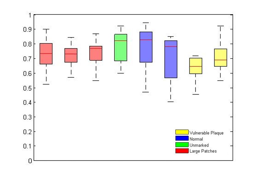
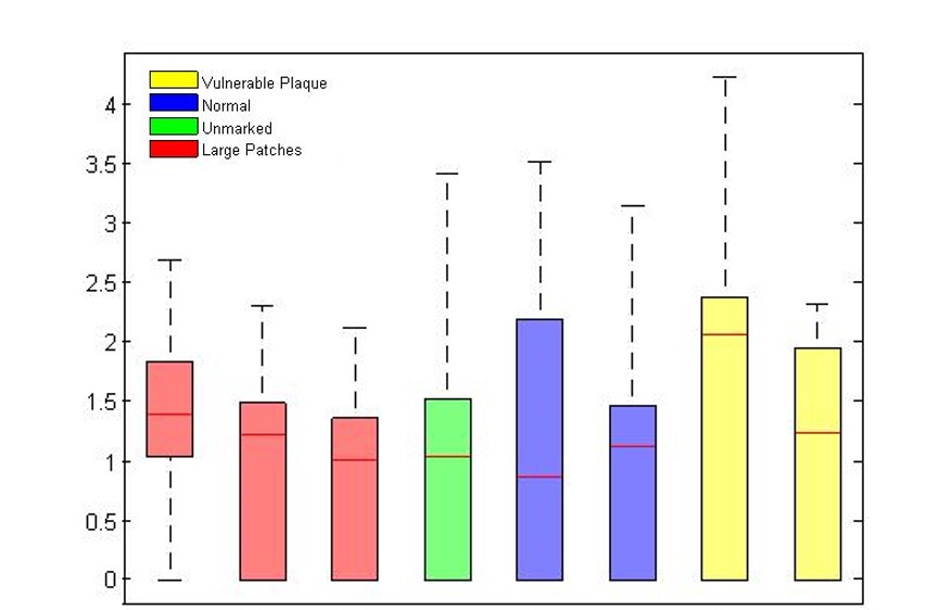
The segmentation results at common carotid artery (CCA), bifurcation, internal carotid artery (ICA) and external carotid artery (ECA) are showed in Figure 1 in which the red line represents the gold standard, and the yellow line represents our results. Figure 2 and Figure 3 exhibited Jaccard coefficient and contour error by comparing the contour segmented automatically with the gold standard.
Discussion
In the eight sets, the Jaccard coefficients (mean: 0.724) were higher than 0.6 and the contour errors (mean: 1.6) were lower than 2.4 pixels. We also found that the patient with vulnerable plaque had low average Jaccard coefficient and high average contour error. This might be associated with image features of vulnerable plaques and parameters selection during iteration.Conclusion
Automatic segmentation based on level set might be an alternative approach to describe the morphology of carotid arteries on TOF MR angiography images.Acknowledgements
None.References
1.Lee SW, Antiga L, Spence JD, et al. Geometry of the carotid bifurcation predicts its exposure to disturbed flow. Stroke. 2008;39:2341-2347.
2. Abdelsamea MM, Gnecco G, Gaber MM, et al. On the Relationship between Variational Level Set-Based and SOM-Based Active Contours. Comput Intell Neurosci. 2015;2015:109029.
3. Li C, Huang R, Ding Z, et al. A level set method for image segmentation in the presence of intensity inhomogeneities with application to MRI. IEEE Trans Image Process. 2011;20:2007-2016.
4. Li C, Xu C, Gui C, et al. Level set evolution without re-initialization: A new variational formulation. Computer Vision and Pattern Recognition, 2005. CVPR 2005. IEEE Computer Society Conference on IEEE Xplore. 2005:430-436.
Figures