3125
Clinical feasibility of Quantitative Susceptibility Mapping with Automatic Uniform Cerebrospinal Fluid Zero Reference1Radiology, Tongji Hospital, Tongji Medical College, Huazhong University of Science and Technology, Wuhan, China, 2Radiolgy, Weill Cornell Medical College, NewYork, NY, United States, 3Biomedical Engineerring, Cornell University, Ithaca, NY, United States
Synopsis
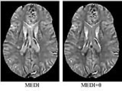
Longitudinal and cross-center studies using conventional quantitative susceptibility mapping (QSM) methods require the choice of a reference tissue, its manual segmentation and subtraction of its average. In this work, we report our initial clinical experience with a fully automated zero-referenced Morphology Enabled Dipole Inversion (MEDI+0) method that uses the ventricular cerebrospinal fluid (CSF) as zero reference in 393 consecutive patients. In 92.62% of cases, excellent agreement of image quality between MEDI+0 and MEDI was observed with high correlation of lesion susceptibility in a combined glioma, ischemic stroke and multiple sclerosis subset of patients.
Background and Purpose: Quantitative susceptibility mapping (QSM) has seen an increasing use in clinical brain studies. Longitudinal and cross-center studies require zero-referencing of the reported susceptibility. In this work, we report on our initial clinical experience with a fully automated zero-referenced Morphology Enabled Dipole Inversion (MEDI+0) in a cohort of 393 consecutive patients. MEDI+0 uses automatically segmented ventricular cerebrospinal fluid (CSF) area within the inversion process to solve the tissue reference problem. Both MEDI+0 and MEDI postprocessing can be performed on the same gradient echo data. The purpose of this study is to evaluate the clinical feasibility of MEDI+0 in different neurological diseases by comparing with MEDI.
Methods: First, 393 consecutive patients imaged during the month of July 2017 on three scanners at our institution were identified for image quality evaluation by a neuroradiologist using five scales: excellent agreement between MEDI+0 and MEDI, good agreement with discrepancy in a very small part of the brain, intermediate agreement with mild disagreement, poor agreement with moderate disagreement, and severe disagreement. Errors in CSF mask computation were also recorded. Next, 20 cases of brain glioma, 21 cases of ischemic stroke and 43 cases (totaling 405 lesions) of multiple sclerosis patients, having all lesions segmented and normal appearing white matter (NAWM) identified, were analyzed using ROI analysis of lesion susceptibility. A linear regression was performed between MEDI+0 and MEDI for these lesion values. Previous studies on MS lesions have used NAWM as reference, therefore, an additional comparison between MEDI and MEDI+0 of susceptibility values using this zero-reference was performed.
Results: In the consecutive 393 cases, 364(92.62%) were scored excellent agreement between MEDI+0 and MEDI, 13 (3.31%) good agreement, 14 (3.56%) intermediate agreement, 2 (0.51%) poor agreement and no severe disagreement. The CSF susceptibility variations in MEDI (range [-28.643, 30.355] ppb, mean 6.253ppb, standard deviation 9.352 ppb) were reduced by MEDI+0 to ([-6.873, 10.742], -0.012, 1.035) ppb. Errors in CSF mask computation occurred in 27 of 393 cases (6.9%), as a result of the presence of a cyst mass, severe edema, vascular malformation, hemorrhage, movement/metal artifact and for a newborn. There was good correlation between MEDI+0 and MEDI when calculating lesion susceptibility of glioma, ischemic stroke, MS referenced to either CSF or NAWM (all P<0.05).
Conclusion: As compared with MEDI, MEDI+0 provides the same image quality, and reliable susceptibility value in different neurological diseases. For lesions near the ventricle, MEDI+0 can better measure the susceptibility without CSF contamination.
Acknowledgements
This work was supported by NIH grant R21EB024366, R21EY027568, R01NS095562, and S10OD021782.References
1. Liu, Z., Spincemaille, P., Yao, Y., Zhang, Y. and Wang, Y., MEDI+0: Morphology enabled dipole inversion with automatic uniform cerebrospinal fluid zero reference for quantitative susceptibility mapping. Magn Reson Med(2017), doi:10.1002/mrm.26946Figures

