3083
Kurtosis and IVIM measurements applied to ischemic stroke diagnosis: an initial experience.1INSERM, UMR 1099, Rennes, France, 2Université de Rennes 1, LTSI, Rennes, France, 3Department of Neuroradiology, Pierre-Zobda-Quitman Hospital, University Hospital of Martinique, Fort-de-France, Martinique, 4Radiology, CH La Palmosa, Menton, France, 5CRLCC, Centre Eugène Marquis, Rennes, France
Synopsis
Diffusional kurtosis imaging (DKI) enables the characterization of non-Gaussian diffusion providing an additional diffusion parameter, the kurtosis (K), that may reflect microstructure heterogeneity. The DKI-IVIM model that incorporates DKI into the IVIM model has been investigated here to assess the feasibility and the potential utility of the DKI-IVIM model for both enhanced diffusion characterization and perfusion measurements in ischemic stroke.
Five stroke patients were enrolled. DKI-IVIM imaging was performed using 8 b-values from 0 to 1500 s/mm2 with a 4 minutes scan duration. IVIM pseudo-diffusion coefficient D*, perfusion fraction f, blood flow-related parameter fD* in addition to the diffusion parameters D (diffusion coefficient) and K were determined in the ischemic lesion and contralateral normal tissue for the stroke patients. Diffusion and perfusion parametric maps were reconstructed.
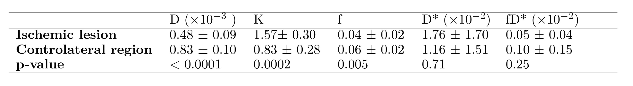
A significant decrease for D (p<0.0001) and increase for K (p=0.0002) in the lesion was observed. The perfusion fraction exhibited a significant decrease in the ischemic regions (p=0.005).
DKI-IVIM model enables for simultaneous cerebral perfusion and enhanced diffusion characterization in an acceptable clinically acquisition time that might improve ischemic stroke diagnosis.
INTRODUCTION
The Intravoxel Incoherent Motion (IVIM) model is a non-invasive method for simultaneous perfusion and diffusion measurements from the diffusion signal1. Due to restricted/hindered diffusion in the intracellular environment, the monoexponential decay that describes water molecules diffusion contribution to the total signal in the conventional model becomes inadequate for b-values exceeding 1000 s/mm2. Diffusional kurtosis imaging (DKI) is an alternative model2 that enables characterization of non-Gaussian diffusion and has been shown to be robust for parameters quantification providing an additional diffusion parameter, the kurtosis (K), that may reflect microstructure heterogeneity. The DKI-IVIM model that incorporates DKI into the IVIM model has been investigated in brain tumors and perfusion studies with encouraging results3,4,5.
The aim of this study was to assess the feasibility and the potential utility of the DKI-IVIM model for both enhanced diffusion characterization and perfusion measurements in ischemic stroke.
METHODS
Five stroke patients were enrolled. DKI-IVIM imaging was performed using 8 b-values from 0 to 1500 s/mm2 chosen with the Cramer-Rao-Lower-Bound optimization approach6 at 1.5T. Sequence parameters were set as following: TR/TE (ms): 3000/75; Matrix size: 256x256; 27 axial slices; NEX=5 with a 4 minutes scan duration. IVIM pseudo-diffusion coefficient D*, perfusion fraction f, blood flow-related parameter fD* in addition to the diffusion parameters D (diffusion coefficient) and K were determined in the ischemic lesion and controlateral normal tissue for the stroke patients from the signal according to the following equation:
$$$\frac{S(b)}{S_0}=f.e^{-bD^*}+(1-f).e^{(-bD+\frac{b^2D^2K}{6})}$$$
with S0 =S (b=0 s/mm2). For each stroke patient, statistical analysis of all DKI-IVIM parameters were performed using the paired Student t-test.
Moreover, both diffusion and perfusion parametric maps were obtained.
RESULTS
For the stroke patients, diffusion signals in both ischemic lesion and normal contralateral region were correctly fitted using the DKI-IVIM model (Figure 1). The paired Student t-test revealed significant differences for all diffusion parameters (Figure 2), with a decrease for D (p<0.0001) and an increase for K (p=0.0002) in the ischemic lesions. The perfusion parameters were reduced with f that exhibits a significant decrease in the ischemic regions (p=0.005), except for D* (p=0.71), due to a high variance on this measurement. The same results could be observed on individual reconstructed parametric maps (Figure 3 and Figure 4).DISCUSSION-CONCLUSION
DKI-IVIM model enables for simultaneous cerebral perfusion and enhanced diffusion characterization in an acceptable clinically acquisition time that might improve ischemic stroke diagnosis. Further improvements are considered for the voxel-by-voxel based analysis.Acknowledgements
No acknowledgement found.References
1. Le Bihan D, Breton E, Lallemand D, Grenier P, Cabanis E, Laval-Jeantet M (1986) MR imaging of intravoxel incoherent motions: application to diffusion and perfusion in neurologic disorders. Radiology 161:401–407.
2. Jensen JH, Helpern JA (2010) MRI quantification of non-Gaussian water diffusion by kurtosis analysis. NMR Biomed 23:698–710.
3. Wu W-C, Yang S-C, Chen Y-F, Tseng H-M, My P-C (2016) Simultaneous assessment of cerebral blood volume and diffusion heterogeneity using hybrid IVIM and DK MR imaging: initial experience with brain tumors. Eur Radiol. doi: 10.1007/s00330-016-4272-z.
4.Pavilla, A, Gambarota, G, Arrigo, A, Mejdoubi, M, Duvauferrier, R, & Saint-Jalmes, H. (2017). Diffusional kurtosis imaging (DKI) incorporation into an intravoxel incoherent motion (IVIM) MR model to measure cerebral hypoperfusion induced by hyperventilation challenge in healthy subjects. Magnetic Resonance Materials in Physics, Biology and Medicine, 1-10.
5. De Luca A, Bertoldo A, Froeling M (2016) Effects of perfusion on DTI and DKI estimates in the skeletal muscle: Effects of Perfusion on DTI and DKI in Muscle. Magn Reson Med. doi: 10.1002/mrm.26373.
6. Leporq B, Saint-Jalmes H, Rabrait C, Pilleul F, Guillaud O, Dumortier J, Scoazec J-Y, Beuf O (2015) Optimization of intra-voxel incoherent motion imaging at 3.0 Tesla for fast liver examination: Optimization of Liver Motion Imaging at 3.0T. J Magn Reson Imaging 41:1209–1217.
Figures
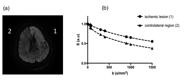
DKI-IVIM analysis on one individual slice of diffusion-weighted images (b = 1000 s/mm2).
(a). Free-hand ROI delineation in the ischemic lesion (marked with “1”) and the corresponding contralateral ROI on normal tissues (“2”).
(b). The corresponding of signal intensity plotted as function of b-value for the two ROIs located in the ischemic lesion and normal contralateral region. Dotted lines represents the fitted curves with the DKI-IVIM model for the lesion and normal regions. The slope related to fD* between b = 0 s/mm2 and b = 300 s/mm2 is decreased for the ischemic region.
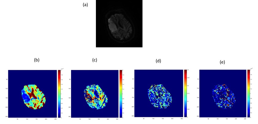
Findings in a 81-year-old patient (female) with stroke.
(a) DWI ( b = 1000 s/mm2), (b) D, and (c) K diffusion parameters, (d) f and (e) fD* perfusion related maps. Restricted diffusion is shown on the D and K maps. The hypoperfusion area is visible on the (d) f and (e) fD* maps.
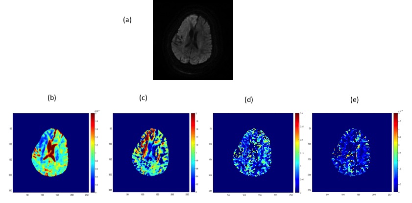
Findings in a 80-year-old patient with stroke(male).
(a) DWI ( b = 1000 s/mm2), (b) D, and (c) K diffusion parameters, (d) f and (e) fD* perfusion related maps. Restricted diffusion is shown on the D and K maps. The hypoperfusion area is visible on the (d) f and (e) fD* maps.