3082
Thalamic nuclei segmentation on dementia using tractography and population-specific priors1Translational Imaging Group, UCL, London, United Kingdom, 2Dementia Research Centre, UCL, London, United Kingdom, 3MRI Unit, Epilepsy Society, Chalfont St Peter, United Kingdom, 4Wellcome EPSRC Centre for Interventional and Surgical Sciences (WEISS), UCL, London, United Kingdom, 5Icometrix, Leuven, Belgium
Synopsis
Thalamic changes have been reported in several neurological disorders, such as Alzheimer's disease and frontotemporal dementia (FTD). As pathologies affect different cortical and subcortical brain regions disproportionally, accurate segmentation of thalamic nuclei can provide relevant insights about brain function and neurological disorders mechanisms. Here, we used a previously developed thalamus parcellation strategy that relies on tractography and population-specific to infer any connectivity changes in presence of FTD. The obtained results were compared against to the ones derived with the commonly used probabilistic tractography pipeline available in FSL.
Introduction
The thalamus is a deep grey matter structure responsible for the propagation of nerve impulses between subcortical and cortical regions in the brain, especially sensory, motor and limbic information. It has different subnuclei that have unique connectivity patterns based on the regions they are connected to. As a result, neurodegeneration affecting any of these structures may lead to secondary degeneration of related brain regions, including the thalamus as shown in Alzheimer’s disease [1] and Frontotemporal dementia (FTD) [2,3]. In this study, we use a previously developed thalamus parcellation pipeline [4] based on diffusion fibre tractography and population-specific priors to infer connectivity changes in the presence of FTD. These findings were compared to the ones obtained with the thalamus tractography-based segmentation pipeline available in FSL [5].Methods
Data processing
A group of 62 participants – 27 controls and 35 FTD patients – was considered in this work. Diffusion-weighted (DW) data consisted of two repeated scans, each with 64 isotropic directions (b-value of 1000 s/mm2) and 4 b-0 volumes, acquired on 1.5T and 3T Philips, Siemens and GE scanners. A T1-weighted MPRAGE image was also obtained with an isotropic voxel size of 1.1 mm3. DW images were corrected for subject-motion, eddy-currents and susceptibility artefacts [6]. T1-weighted images were bias-field corrected, followed by brain parcellation into a total of 143 regions [7]. Finally, affine intra-subject registration was carried out between DW (float) and T1 (target) spaces [8].
Tractography
Six cortical masks (prefrontal, motor, sensory, parietal, occipital and temporal), created as aggregates from 143 regions, and one thalamus mask were selected per hemisphere from the original brain parcellation, and then propagated to the DW space by using the inverse of the previous computed transformation. Fibre orientation distributions on DW data were estimated using constrained spherical deconvolution (CSD) [9]. Finally, probabilistic tractography was performed using iFOD2 algorithm [10] and structural connectivity was reconstructed between every seed voxel in the thalamus and each cortical mask separately. In total, six connectivity maps were derived, representing the probability for each thalamic voxel to be connected to a certain cortical region.
Thalamus parcellation pipeline
Thalamic nuclei segmentation was obtained by using a previously proposed method (PM) [3] that relies on tractography and population-specific thalamic nuclei priors. Briefly, this iterative method involves the construction of a group mean by performing group-wise registration of T1 and FA images of all participants. The connectivity maps of each subject are then mapped to the common space and averaged to derive the population-specific thalamic nuclei priors. These priors are propagated back to the individual’s space and used to infer new connectivity maps.
Application to Frontal Temporal Dementia
Thalamus parcellations were derived, both for normal controls and FTD patients, using the PM and FSL frameworks. The resulting six connectivity-defined regions (CDRs) from each strategy were compared between the two groups by assessing any differences in their normalised volumes to the total intracranial volume (TIV).
Results
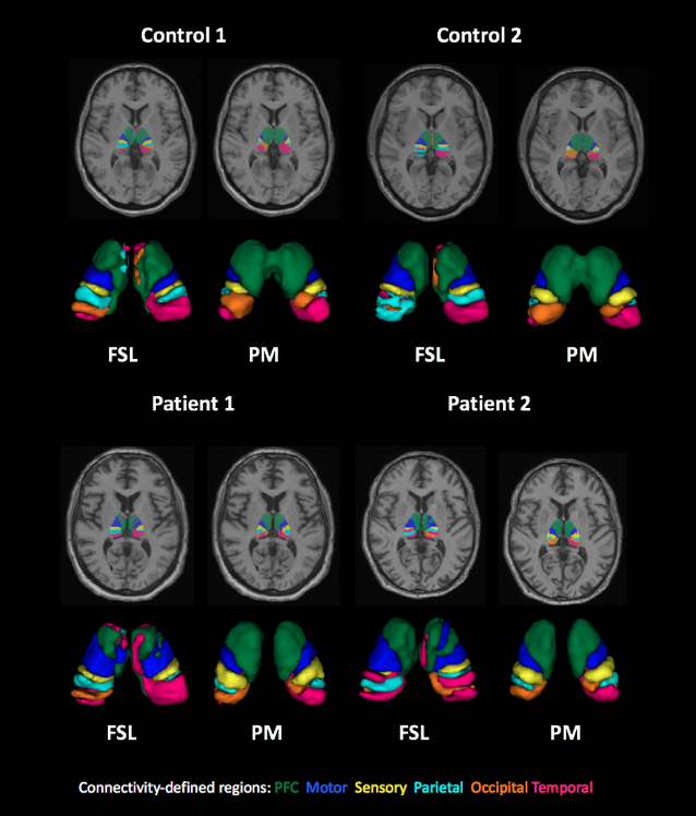
Thalamus connectivity-based segmentations obtained with PM and FSL strategies are displayed in Fig. 1. PM has led to parcellations that are in better agreement with known anatomy and with less fragmented clusters, both for healthy subjects and FTD patients.
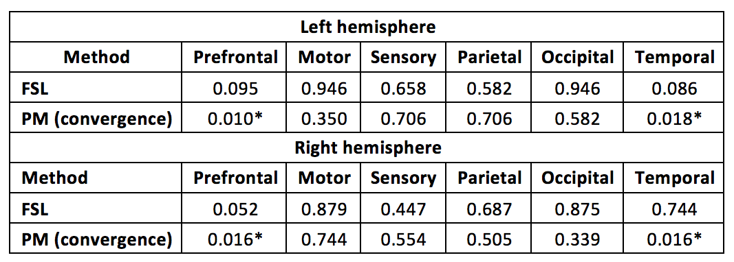
Volume comparisons of CDRs between controls and FTD patients were done independently for each hemisphere and method using the Mann-Whitney test, followed by multiple-comparison correction with false discovery rate (FDR) strategy. Table 1 describes the FDR adjusted p-values derived for each method and CDR on left and right hemispheres. Significant volume differences were observed on prefrontal and temporal CDRs with the PM, whereas no differences were detected with FSL. Previous FTD studies have reported volume differences on prefrontal and temporal cortices, which it is consistent with the results obtained with PM for the off-site connected thalamic nuclei.
Discussion
The performance of tractography in each subject’s native space and integration of population-specific thalamic nuclei priors has led to more biological plausible thalamus parcellations, in the sense that PM was sensitive to connectivity differences between controls and FTD patients, which are in agreement with previous FTD studies [2,3].Acknowledgements
CS is funded by the Engineering and Physical Sciences Research Council (ESPRC) and Icometrix industrial partner.References
[1] Jong et al., Brain, 2008; [2] Rohrer JD et al., Lancet Neurology, 2015; [3] Cardenas VA et al., Archives Neurology, 2007; [4] Semedo C et al., ISMRM, 2017; [5] Behrens et al., NeuroImage, 2007; [6] Clarkson MJ et al., Int J Comput Assist Radiol Surg., 2015 [7] Cardoso et al., IEEE TMI, 2015; [8] Modat et al., Computer Methods and Programs in Biomedicine, 2010; [9] Tournier et al., NeuroImage, 2007; [10] Tournier et al., ISMRM, 2010.Figures