3069
Monitoring effect of rapamycin on pyruvate metabolism in SCC tumor using hyperpolarized 13C-MRI1National Cancer Institute, Bethesda, MD, United States, 2Hokkaido University School of Engineering, Sapporo, Japan, 3National Institutes for Quantum and Radiological Science and Technology, Chiba, Japan, 4Gifu University School of Medicine, Gifu, Japan, 5National Institute of Neurological Disorder and Stroke, Bethesda, MD, United States
Synopsis
Effect of an mTOR inhibitor rapamycin on pyruvate metabolism in squamous cell carcinoma (SCC) xenografts was investigated using hyperpolarized 13C-MRI. [1-13C]lactate to [1-13C]pyruvate ratio (Lac/Pyr) in the SCC tumors increased as tumor grew in non-treated control mice, whereas it significantly dropped after 2 days of the rapamycin treatment. Inhibition of mTOR caused a drop of LDH protein level and the activity in the SCC tumor, and perfusion in the tumor was improved by the rapamycin treatment. Lac/Pyr monitored using hyperpolarized 13C-MRI would become a useful marker for tumor response to mTOR inhibitors.
Introduction
Mammalian target of rapamycin (mTOR) is a protein kinase that is centrally involved in the control of cancer cell metabolism, growth, and proliferation, and therefore has become an attractive therapeutic target. Rapamycin is an inhibitor of mTOR, and inhibition of mTOR by rapamycin causes diverse effects on tumor microenvironment, such as oxygenation and energy metabolism. Such changes can serve as markers to assess tumor response to rapamycin therapy. We investigated effects of rapamycin on pyruvate metabolism to lactate in squamous cell carcinoma (SCC) xenograft using hyperpolarized 13C-MRI. Lactate dehydrogenase (LDH) is downstream protein of mTOR and expected to be affected by rapamycin treatments.Methods
SCC cells (5x10⁵ cells) were implanted subcutaneously into a right hind leg of female C3H mice. Treatment with rapamycin and MRI scans were started 8 days after the implantation of the SCC tumor at that time the size of SCC tumors was around 600 mm3. [1-13C]pyruvate containing 15 mM OX063 was polarized for approximately 1 hour using a hyperpolarizer (HyperSense, Oxford Instruments), and the hyperpolarized [1-13C]pyruvate (300 µL of 96 mM solution) was injected intravenously to the tumor-bearing mice. The MRI scans were carried out using a 4.7 T scanner (Bruker Bio-Spin MRI GmbH) with a 17 mm home-built 13C solenoid coil placed inside of a saddle coil for 1H.Results
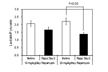
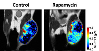
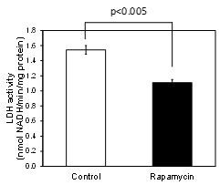
To confirm mTOR inhibition causes a drop of LDH activity in SCC tumor cells, LDH activity and LDH protein level in SCC cells were assessed with and without rapamycin (100 nM, 48 h). LDH activity in the cell lysates estimated from NADH consumption in the presence of pyruvate was significantly smaller in rapamycin treated cells compared to non-treated control cells (Figure 1). The LDH protein level in rapamycin treated cells also dropped to 49.1±8.6 % of the non-treated control SCC cells. Pyruvate metabolism to lactate in SCC tumor xenograft was evaluated using 13C-MR spectroscopy with hyperpolarized [1-13C]pyruvate. Signals of [1-13C]pyruvate and [1-13C]lactate were detected in the SCC tumor immediately after hyperpolarized [1-13C]pyruvate injection, indicating that exogenously injected pyruvate was quickly converted to lactate by LDH catalyzed reaction in the SCC tumor. To assess rapamycin effect on lactate formation, lactate to pyruvate ratio (Lac/Pyr) was calculated from area under the curve of signal intensity curves of [1-13C]lactate and [1-13C]pyruvate. In non-treated SCC tumors, Lac/Pyr increased as tumor grew from 1.62±0.12 on day 8 to 2.26±0.23 on day 10. On the other hand, Lac/Pyr was significantly dropped by rapamycin treatments from 1.63±0.19 on day 8 (before rapamycin treatments) to 1.07±0.07 on day 10 (after 2 days rapamycin treatments). Then, chemical shift images in SCC tumors were acquired 30-50 sec after hyperpolarized [1-13C] injection (Figure 2). Lac/Pyr in the tumor region calculated from chemical shift images also showed similar behavior to the 13C-MRS results; lactate formation dropped by rapamycin in a dose dependent manner (Figure 3). We also carried out DCE-MRI with gadolinium-DTPA to evaluate changes of perfusion in the SCC tumor by rapamycin treatments. The Gd-DTPA uptake was significantly higher in the rapamycin treated group compared to the non-treated control group (Figure 4), indicating tumor perfusion was improved by the rapamycin treatment.Conclusion
Pyruvate metabolism to lactate was dropped by rapamycin treatments in SCC tumor, and both the drop of LDH activity and the improvement of tumor perfusion contributed to the Lac/Pyr drop. The results suggest that Lac/Pyr monitored with hyperpolarized 13C-MRI could become a useful marker for evaluating early tumor response to mTOR inhibitors.Acknowledgements
No acknowledgement found.References
No reference found.Figures
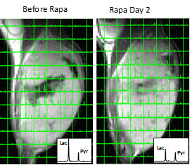
13C-chemical shift images and averaged spectra in a SCC tumor obtained before and after 2 days of rapamycin treatments (20 mg/kg/day).