3046
A proof-of-concept study on the quantification of gene expression levels with doxycycline-inducible MR reporter gene1MR Core, Asan medical Center, Seoul, Republic of Korea, 2GangNeung Asan Medical Center, GangNeung, Republic of Korea, 3Radiology, Asan medical Center, Seoul, Republic of Korea
Synopsis
Recent research on MR reporter genes has demonstrated their potential for use in transgene expression monitoring. We have conducted a preliminary study on the development of a new MRI reporter system [organic anion transporting polypeptide (OATP) 1B1] that can analyze gene expression level using MR reporter genes. By establishing doxycycline-inducible cell line, we observed T1 shortening on MRI, which indicated increased expression level of the OATP1B1 gene. A strong correlation was observed between conventional methods for measurement of gene expression and rrT1 of MR imaging. In this study, we provide preliminary evidence of the potential application of MRI to determine gene expression.
Introduction
Since the pioneering phase of MR reporter genes, several advances have been made in the field of MR reporter gene imaging.1-3 MR reporter genes may be used for transgene expression monitoring; however, only a few studies have used MR reporter for the quantification of gene expression levels.4-6 In this study, we built a doxycycline-inducible organic anion transporting peptide 1B1 (OATP1B1) expression system and investigated whether the doxycycline-inducible OATP1B1 MR reporter gene can be used for the quantification of gene expression levels.Methods
Tet-On OATP System: We
used Tet-on Advanced Inducible Gene Expression System (Clontech Lab., USA) and
constructed a recombinant vector containing human OATP1B1 cDNA (pTRE-OATP
plasmid) (Fig. 1A, 1B).
Tet-HEK1B1 cell line establishment: To
establish Tet-HEK1B1 cells expressing the OATP1B1 gene by doxycycline
induction, pTRE-OATP plasmid was transfected into HEK cells; cells were then
treated with G418 (1 mg/mL) and hygromycin (0.5 mg/mL) for 3–4 weeks to sort
out cells that expressed pTet-On and pTRE-OATP. Cells were then treated with doxycycline
for 24 h (10–300 ng/mL).
Validation of OATP1B1 expression: For western blot, cells were lysed with RIPA buffer. Lysates were run on 10% SDS PAGE gel and transferred on to nitrocellulose membranes. GFP and OATP1B1 antibodies were used to blot the proteins. Expression of GFP (indicating the transcription of pTRE-OATP) was confirmed using a confocal microscope (LSM 780, Zeiss, Germany).
Preparation of cell phantom: Cells were grown to 70% confluence and incubated with/without gadoxetate (0.5 mM) in a growth medium for 20 h. After incubation, cells were harvested and washed twice in phosphate-buffered saline and lysed with RIPA buffer. Pellet was collected in a 0.2-mL tube for in vitro MR phantom study.
In vitro MR phantom and analyses: MR imaging was performed on a 9.4 T/160-mm MR system (Agilent Inc.). T1 sequences were obtained with a fast spin-echo inversion-recovery: TR/TE = 4700/7.4 ms; TI set = 20, 50, 100, 200, 400, 1000, 2000, 4000 ms; ETL = 8; segment = 12; and resolution = 0.26 mm. The reduction rate of T1 shortening time (rrT1) was calculated using the following equation:
rrT1 (%) = [(average Gd 0mM − 0.5 mM)/ave Gd 0 mM] * 100
Results
For the validation of the OATP1B1 inducible system, we performed western blot analysis, which showed expression of OATP1B1 in the Tet-HEK1B1 cells after doxycycline treatment (100 ng/mL; Fig. 1C). Results of T1 mapping analysis showed that rrT1 for doxycycline-induced Tet-HEK1B1 cells was significantly larger than that for HEK293 cells (Figs. 2B–2D). After Tet-HEK1B1 cell line establishment, we performed doxycycline dose-dependent OATP1B1 induction experiments with Tet-HEK1B1 cell line. A gradual increase in OATP1B1 gene expression level was observed on western blot, and GFP-positive signals also increased with doxycycline dose increase (Figs. 3A, 3B). We then analyzed T1 mapping images of Tet-HEK1B1 cell phantoms treated with different doxycycline doses; we found that Tet-HEK1B1 cells treated with high-dose (100 ng/mL) doxycycline exhibited greater T1 shortening effect than those treated with low-dose doxycycline (10 ng/mL) (Fig. 3D). Moreover, on linear regression analysis, we observed a positive correlation between OATP1B1 protein levels and rrT1 values of MR images.Discussion
Although our results suggest that the MR reporter can be used for the quantification of gene expression levels, some limitations and concerns need to be addressed. For a meaningful application of MR reporter as a gene expression quantification tool, new reporter systems with enhanced sensitivity are needed. Compared with the average transcription levels of several clinically and biologically important genes, the expression level of doxycycline-induced gene is much higher as the Tet-system is primarily used to study gene function by overexpression. Therefore, in addition to sensitivity enhancement, validation of the transcription level of the genes of interest in normal or abnormal physiological settings is required. Moreover, investigation of the biological effects of MR reporter expressions on cell physiology is also necessary.Conclusion
In conclusion, our results provide preliminary proof of the potential use of MRI for the quantification of gene expression levels. However, further research is required for meaningful application of MRI reporters in biological and clinical investigations.Acknowledgements
This study was supported by grants from the Basic Science Research Program through the National Research Foundation of Korea [NRF-2015R1C1A1A02036526] and the Korea Health Technology R&D Project through the Korea Health Industry Development Institute [HI14C1090], funded by the Ministry of Health & Welfare, Republic of Korea.References
1. Gilad, A.A., et al., MRI reporter genes. J Nucl Med, 2008. 49(12): p. 1905-8.
2. Gilad, A.A., et al., Developing MR reporter genes: promises and pitfalls. NMR Biomed, 2007. 20(3): p. 275-90.
3. Gilad, A.A. and M.G. Shapiro, Molecular Imaging in Synthetic Biology, and Synthetic Biology in Molecular Imaging. Mol Imaging Biol, 2017. 19(3): p. 373-378.
4. Wang, K., et al., MR reporter gene imaging of endostatin expression and therapy. Mol Imaging Biol, 2010. 12(5): p. 520-9.
5. Patrick, P.S., et al., Dual-modality gene reporter for in vivo imaging. Proc Natl Acad Sci U S A, 2014. 111(1): p. 415-20.
6. Gilad, A.A., et al., Artificial reporter gene providing MRI contrast based on proton exchange. Nat Biotechnol, 2007. 25(2): p. 217-9.
Figures
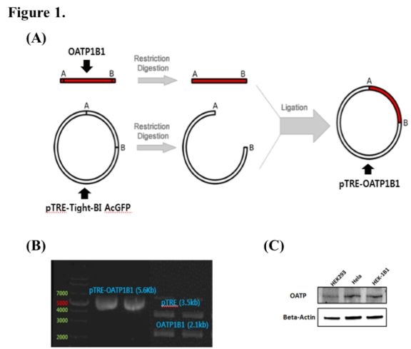
Fig. 1. pTRE-OATP plasmid and stable cell line establishment.
(A) Schematic of pTRE-OATP plasmid generation by gene cloning.
(B) Confirmation of pTRE-OATP plasmid by enzyme cutting.
(C) Western blot analysis of HEK293 (OATP-negative), HeLa (OATP-positive), and HEK-1B1 (pTRE-OATP plasmid-transfected) cell lines.
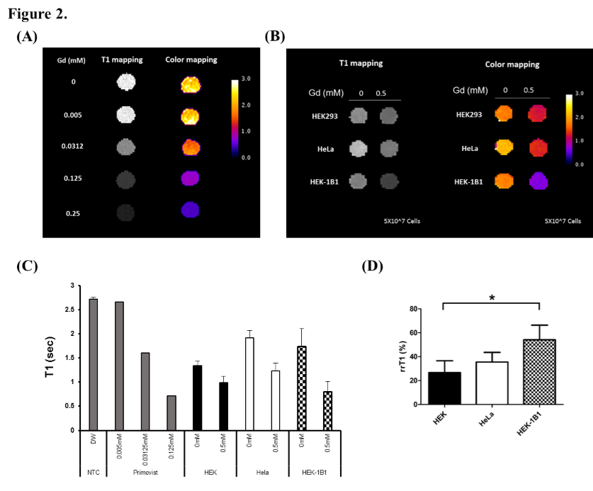
Fig. 2. Evaluation of the OATP1B1 gene expression ON/OFF by MRI.
(A) Image of T1 mapping of Gadoxetate (Gd) titration.
(B) Image of T1 mapping of HEK293 (OATP-negative), HeLa (OATP-positive) and HEK-1B1 (pTRE-OATP plasmid-transfected) cell lines.
(C) T1 results of (A) and (B).
(D) rrT1 calculation of (B) (*p < 0.05; two-tailed Student’s t-test).
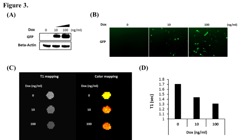
Fig. 3. Evaluation of the OATP1B1 gene expression level by MRI.
(A) Western blot analysis of HEK-1B1 (pTRE-OATP plasmid-transfected) cell lines treated with low and high concentrations of doxycycline.
(B) Expression of OATP1B1 (by GFP signal) using confocal microscopy.
(C) Image of T1 mapping and color mapping of HEK-1B1 (pTRE-OATP plasmid-transfected) cell lines treated with low and high concentrations of doxycycline.
(D) T1 results of (C).