3038
Fluorine-19 MRI hot-spot imaging of lung metastasis in rodents1Radiology, University of California, San Diego, CA, United States
Synopsis
Lung cancer is the leading cause of cancer deaths. Safe and specific MRI probes are needed to enable early detection of lesion presence and therapeutic response. Injected PFC nanoemulsion, taken up by tumor associated macrophages (TAMs), can be used as a biomarker to detect metastases using 19F MRI. In a metastatic lung cancer mouse model, we show that PFC is effectively taken up by TAMs and vividly displays lung metastasis using 19F MRI. Validation assays using in vivo bioluminescence and histology support the MRI findings. Overall, 19F hot-spot imaging offers a highly-specific marker of tumor burden in lung parenchyma.
INTRODUCTION
Lung cancer is the second most common cancer in men and women, and the leading cause of cancer deaths. Current standards for detection include tissue biopsy, an invasive procedure for which the detection of cancer lesions is highly dependent on the biopsy location, and chest CT scans which expose patients to potentially harmful radiation and for which soft tissue contrast is generally low, making detection of small metastatic lesions challenging. Safe and specific MRI probes are needed to enable early detection of lesion presence and therapeutic response. As a hallmark of cancer, inflammation is a key component in the function of the tumor microenvironment. Perfluorocarbon (PFC) nanoemulsion injected intravenously is taken up by the reticuloendothelial system which is comprised of mononuclear, phagocytic cells, including macrophages. PFC uptake by tumor-associated macrophages (TAMs) can therefore be used as an in vivo biomarker for lesion detection1,2. In a metastatic lung cancer mouse model, we show that PFC is effectively taken up by TAMs and vividly displays lung metastasis using 19F MRI. Validation assays using in vivo bioluminescence (BLI) and histology support the MRI findings. Preliminary data suggests that 19F hot-spot imaging offers a highly-specific marker of tumor burden in lung parenchyma.METHODS
SCID beige female mice (N=10) of 6-7 week age were inoculated via tail vein with 5x10⁶ A549-luc-C8 cells (PerkinElmer). The presence, location and progression of tumors was initially assessed by BLI weekly for three weeks. BLI scans were acquired using an IVIS Spectrum (PerkinElmer) in vivo imaging system 10 min post-IP injection of luciferin (150 mg/kg) using large pixel binning with F/Stop=1, and the scan time was scaled to acquire 2-3x10⁵ total counts. Seventy percent (7/10) mice developed lung metastases by three weeks post-cell injection, five of which were selected for injection with PFC nanoemulsion (~150 nm mean particle size, 30% v/v, V-Sense, Celsense) 48 hour prior to MRI. Mice were anesthetized with 1-2% isoflurane in oxygen and kept warm using a water-heated imaging bed. 19F RARE MRI scans were acquired using a Bruker 11.7T system with TR/TE=825/18 ms, RARE factor 8, 3×3 cm FOV, 32x32 matrix and 2 mm-thick axial slices covering lung and liver regions. The number of averages was scaled to acquire a 30-45 min scan. Co-registered 1H RARE scans were acquired with TR/TE= 950/18 ms, RARE factor 8, 3×3 cm FOV, 192x192 matrix and 1 mm-thick axial and coronal slices. The number of slices was doubled to obtain the same coverage as the 19F RARE scan. At the experimental endpoint animals were sacked for histology. Images were analyzed using Living Image 4.1 for BLI and VivoQuant 3.5 (Invicro) for 19F/1H composite image rendering.RESULTS/DISCUSSION
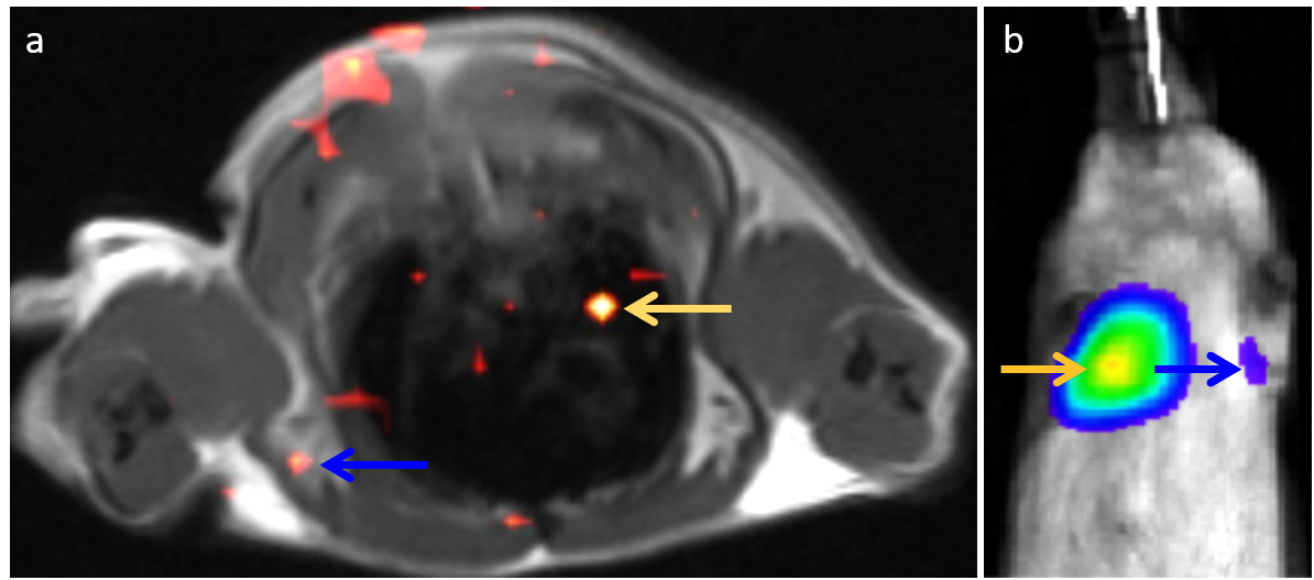
The 19F MRI data show localized uptake of PFC in lung, presumably from A549-luc-C8 lung metastases; representative data is displayed in Fig. 1a. The relative location of metastases was confirmed by BLI in Fig. 1b, however, anatomical MRI scans were not able to resolve the lesions (not shown). Interestingly, in some cases, the 19F MRI shows distinct signals throughout the lungs indicating multiple lesions compared to BLI detection which shows a single tumor signal in the thoracic region. The intensity of 19F signals also correlates to BLI signal intensity, suggesting increased pro-inflammatory activity in larger tumor lesions. The results demonstrate the feasibility of PFC as a biomarker for 19F MRI detection of lung metastases. The inflammatory sensitivity of this biomarker also suggests a potential use in immuno-oncology. Therapies in these studies focus on modulating immune response to treat cancer, and thus a biomarker for tumor-related inflammation may be useful in early evaluation of treatment response. 19F MRI additionally overcomes challenges associated with 1H MRI’s low specificity and resolution limitations due to motion artifacts when imaging the thoracic region.Acknowledgements
This work was supported by the NIH grants R01-EB017271, R01 CA139579, R01 EB024015 and CIRM grant LA1-C12-06919.References
1. Khurana A, Chapelin F, Xu H, Acevedo JR, Molinolo A, Nguyen Q, Ahrens ET. Visualization of macrophage recruitment in head and neck carcinoma model using fluorine-19 magnetic resonance imaging. Magn Reson Med. 2017 Jul 26. doi: 10.1002/mrm.26854.
2. Makela A, Gaudet J, Foster P. Quantifying tumor associated macrophages in breast cancer: a comparison of iron and fluorine-based MRI cell tracking. Sci Rep. 2017; 7: 42109. doi: 10.1038/srep42109.
Figures