3027
A Motion Correction Method Based on Navigator for Simultaneous PET/MR abdominal ImagingKe Meng1,2,3, Lingzhi Hu4,5, and Qun Chen5
1Shanghai Advanced Research Institute, Shanghai, China, 2University of Chinese Academy of Sciences, Beijing, China, 3Shanghai Tech University, Shanghai, China, 4UIH America Inc, Houston, TX, United States, 5United Imaging Healthcare, Shanghai, China
Synopsis
The integrated PET/MR combines the advantages of functional imaging device PET and high resolution high contrast MRI, simultaneously acquiring PET and MR images at the same position, improving image fusion accuracy. However, respiratory motion during abdominal imaging causes notorious motion artifact in the MRI images and blurring the PET images. A PET/MR motion correction method based on real-time 2D excitation navigator has been implemented and evaluated. Phantom and human imaging result implies that this technique can precisely acquire object motion and effectively eliminate motion blurring. Without additional operation and device, it offers a simple and cost-down way for clinical use.
Target audience
Clinicians
and medical researchers who are interested in advanced applications of next
generation PET/MR systemPurpose
The
integrated PET/MR combines the advantages of functional imaging device positron
emission tomography and high resolution high contrast magnetic resonance
imaging, simultaneously acquiring PET and MR images at the same position,
improving image fusion accuracy. However, respiratory motion during abdominal
imaging causes notorious motion artifact in the MRI images and blurring the PET
images. To improve diagnosis accuracy, motion blurring needs to be eliminated or
reduced in PET/MR scan. In this work, a PET/MR motion correction method based
on real-time 2D excitation navigator has been implemented and assessed on phantom
and clinical data. Methods
A
motion correction strategy contains two steps: (1) extracting precise motion
vector during MRI acquisition; (2) correcting PET list mode data according to
the motion vector. All experiments were performed on a simultaneous TOF PET/MR
scanner (uPMR790, United Imaging Healthcare, Shanghai, China). A pencil beam
navigator excited by a 2D RF pulse was positioned along the motion direction
and the projection profile was obtained by the Fourier transform of the
measured signal. By utilizing an edge detection algorithm, the respiratory
motion vector can be extracted. Then the estimated motion is deployed to
prospectively gating the MR data acquisition and retrospectively re-binning
PET data. To verify this approach, three point-sources was positioned in a
shaddock. Bed drives shaddock moving back and forth to imitate a respiratory
motion. In the human scans, navigator was placed across the right
hemi-diaphragm along the superior-inferior direction and respiratory belt was
placed around the upper abdomen to acquire respiratory signals for contrast. Simultaneous
PET data acquiring and navigator interleaved with FSE sequence are implemented
in uPMR 790 PET/MR system (United Imaging Healthcare, Shanghai, China).Results
Navigator
accurately tracked the abdominal motion and the motion vector extracted by edge
detection algorithm well delineated the motion (Figure2). Motion blurring in the
PET image was significantly reduced after correction (Figure 3). Signal to
noise ratio (SNR), ghost to image ratio (GIR) of MR images and FWHM values of
lesions in PET images has been evaluated quantitatively in phantom. SNR
increased from 163.89 to 662.23 whereas GIR reduced from 24.87% to 2.94%,
suggesting a significant improvement compared to the one without motion
correction. In human scan, there is no significant difference (P>0.8) of the
image quality that obtained with navigator (SNR = 89.35, GIR= 7.93%) and respiratory belt (SNR = 91.05, GIR
= 8.15%).Discussion
Respiratory
motion is the main cause of artifacts in abdominal imaging. A motion correction
strategy based on navigator for simultaneous PET/MR has been developed and
evaluated. Phantom and human imaging result implies that this technique can
precisely acquire object motion and effectively eliminate motion blurring. Conclusions
We
have successfully implemented and evaluated a respiratory gating sequence for
abdominal imaging in a simultaneous PET/MR. Based on the qualitative and
quantitative analysis of phantom and human data, we can conclude that navigator
provides a respiratory monitor which is suitable for PET/MR motion correction
in abdomen imaging. Reducing motion artifacts and improving diagnosis accuracy,
without additional operation and device, it offers a simple and cost-down way
for clinical use. Acknowledgements
No acknowledgement found.References
No reference found.Figures

Figure1. Shaddock in uPMR790
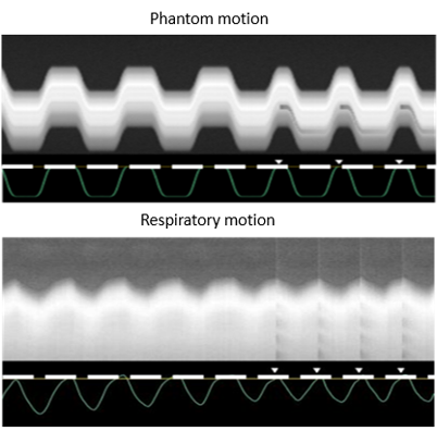
Figure 2. Motion information extracted by navigator

Figure 3. Motion correction results on shaddock
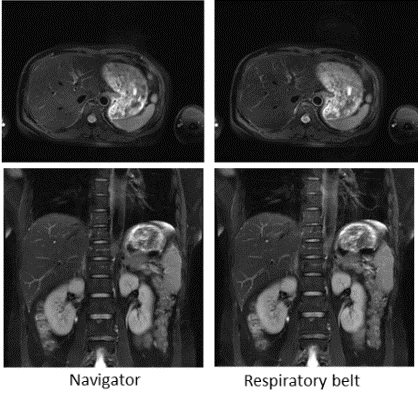
Figure 4. Navigator corrected MR images and a comparison of respiratory belt

Figure 5. Abdomen image after correction