3011
Association between Incompleteness of Circle of Willis and Carotid Vulenrable Atherosclerotic Plaques: A CARE-II StudyChangwu Zhou1, Chun Yuan2,3, Wei Wang1, Cheng Li4, and Xihai Zhao3
1Radiology, The affiliated hospital of YangZhou University, YangZhou, China, 2Department of Radiology, University of Washington, Washington, American Samoa, 3Center for Biomedical Imaging Research, Department of Biomedical Engineering, Tsinghua University School of Medicine, Beijing, China, 4Radiology, Department of Radiology, Zhongda Hospital, Medical School of Southeast University, Nanjing, China
Synopsis
The circle of Willis (COW) is an important intracranocervical collateral circulation system. We hypothesized that the integrity of COW may affect the characteristics of carotid plaques by influencing carotid hemodynamics. This study investigated the relationship between incompleteness of COW and the compositional features of atherosclerotic plaques in carotid arteries. We found that the incompleteness of circle of Willis is associated with vulnerability of carotid artery atherosclerotic plaques. Our findings suggest that integrity of circle of Willis may play a role in occurence of high risk plaque features, particularly intraplaque hemorrhage and fibrous cap rupture.
Introduction
The circle of Willis (COW) is an important intracranocervical collateral circulation system [1]. COW can maintain adequate blood flow through its potential redistribution function according to the anatomical morphology of COW, especially in patients with cerebrovascular diseases [2-5]. We hypothesized that the integrity of COW may affect the characteristics of carotid plaques by influencing carotid hemodynamics. This study sought to investigate the relationship between incompleteness of COW and the compositional features of atherosclerotic plaques in carotid arteries.Methods
Study sample: Four hundred and eighty-two symptomatic patients (mean age 61.2±10.4 years, 315 males) enrolled in a multicentre study of A Chinese Atherosclerosis Risk Evaluation II (CARE-II, NCT02017756) were recruited in this study. All patients underwent a routine 3D TOF MR angiography (MRA) for intracranial arteries and 2D multicontrast MR vessel wall imaging for carotid arteries on a 3.0T MR scanner (Philips Achieva TX) with a 8-channel dedicated carotid coil. The imaging parameters for TOF MRA are as follows: 3D TOF: fast field echo, repeat time/echo time 25/3.5 ms, flip angle 20°, field of view 4.5×20×20 cm3, and spatial resolution 0.7×0.7×1.4 mm3. Carotid multicontrast MR vessel wall imaging was performed using a standard protocol including TOF, T1W-QIR, T2-MDIR, and MP-RAGE with published parameters [6]. The study protocol was approved by institutional review board and written consent form was obtained from each subject. Image review: The circle of Willis (COW) was divided into four parts anatomically (Fig. 1): anterior COW (A-COW), posterior COW (P-COW), left COW (L-COW) and right COW (R-COW). The integrity of COW was evaluated. The absence of any structure on MRA images in each of the four parts was considered as incompleteness of COW. For example, the absence of anterior communicating artery (or left anterior cerebral artery: ACA-A1 or right ACA-A1) was considered as incompleteness of A-COW. Carotid MR images were reviewed by two radiologists with >5 years’ experience in cardiovascular MR imaging using “CASCADE” (UW, Seattle, USA). Presence/absence of plaque components, such as calcification, lipid-rich necrotic core, intraplaque haemorrhage (IPH), and fibrous cap rupture was determined. Statistical analysis: The prevalence of incompleteness of COW according to different plaque compositions in carotid arteries was calculated and compared using Chi-square analysis.Results
Of 482 patients, 421 (87.3%) had the incompleteness in COW. The prevalence of incompleteness was significantly higher in P-COW than A-COW (79.7% vs. 40.7%, p<0.001). There was no significant difference between L-COW and R-COW (77.0% vs. 76.6%, p=0.939). For carotid plaque characteristics, 48.1%, 73.0%, 15.4% and 8.1% patients were found to have calcification, LRNC, IPH, and fibrous cap rupture, respectively. The prevalence of incompleteness in A-COW was significantly higher in patients with IPH than those without IPH (52.7% vs. 38.5%, p=0.022) (Table 1, Fig. 2), especially the absence of anterior communicating artery (69.2%, p=0.001). The prevalence of incompleteness in P-COW was significantly higher in patients with FCR than those without FCR (92.3% vs. 78.6%, p=0.041, Table 1). No significant differences were found in prevalence of incompleteness in COW between patients with and without calcification and LRNC (all p>0.05, Table 1).Discusion and Conclusion
The incompleteness of circle of Willis is associated with vulnerability of carotid artery atherosclerotic plaques. Our findings suggest that integrity of circle of Willis may play a role in occurence of high risk plaque features, particularly intraplaque hemorrhage and fibrous cap rupture.Acknowledgements
We present this study on behalf of CARE-II study collaborators.References
[1] Sallustio F, et al. Stroke. 2008;39:1894-7. [2] Tanaka H, et al. AJNR. 2006;27:1770-5. [3] Liebeskind D. Stroke. 2003;34:2279-84. [4] Rutgers DR, et al. Stroke. 2004;35:1345-9. [5] Hendeikse J, et al. Radiology. 2005;235:184-9. [6] Xihai Zhao, et al. SVN. 2017.Figures
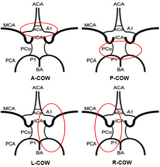
Fig1. The four parts
of COW
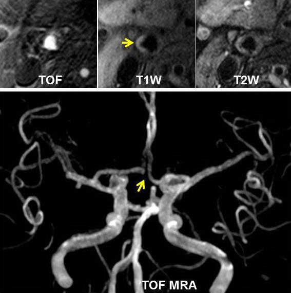
Fig2. IPH was found in right carotid
artery (yellow arrow on T1W). The anterior communicating artery was absent on
TOF MRA image in the same patient.
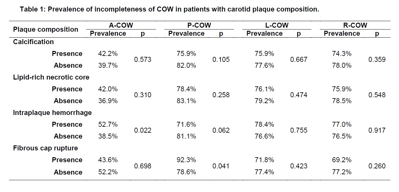
Table1 : Prevalence of incompleteness
of COW in patients with carotid plaque composition.