3002
Reproducibility of Simultaneous Intracranial and Extracranial Arterial Vessel Wall MR Imaging based on T1 weighted DANTE-SPACE1Shenzhen Institutes of Advanced Technology, Chinese Academy of Sciences, ShenZhen, China
Synopsis
Intracranial and extracranial atherosclerotic disease are major causes of ischemic stroke. Recently, an improved DANTE-prepared 3D variable-flip-angle turbo spin echo (SPACE) imaging method was developed for high resolution simultaneously imaging of intracranial and extracranial arterial vessel wall with enhanced cerebrospinal fluid suppression. The purpose of this study was to evaluate the scan-rescan, intra-and inter-observer reproducibility when using the method for comprehensive assessment of intracranial and extracranial vessel wall morphology. In conclusion, the improved 3D simultaneous vessel wall imaging technique provided good to excellent reproducibility for intracranial and extracranial arterial vessel wall measurements.
Introduction
Intracranial and extracranial atherosclerotic disease are major causes of ischemic stroke. Magnetic resonance (MR) vessel wall imaging with large spatial coverage from common carotid artery to distal intracranial artery based on three dimensional (3D) variable flip angle turbo spin echo (SPACE) combined with Delay Alternating with Nutation for Tailored Excitation (DANTE) module (referred as DANTE-SPACE) was proposed to simultaneously image intracranial and extracranial arterial vessel wall.1 However, evaluation of cerebrospinal fluid (CSF) suppression in this method was limited to the cervical spinal cord region, and the low spatial resolution made depiction of distal middle cerebral arteries (MCA) difficult. Currently, an improved DANTE-SPACE method was developed to uniform CSF suppression around the MCA without affecting blood suppression at the carotid region, and which would achieve an isotropic spatial resolution of 0.55 mm.2 To meet the clinical needs, this imaging technique should be reproducible. The purpose of this study was to evaluate the scan-rescan, intra-and inter-observer reproducibility when using the method for comprehensive assessment of intracranial and extracranial vessel wall morphology.Materials and Methods
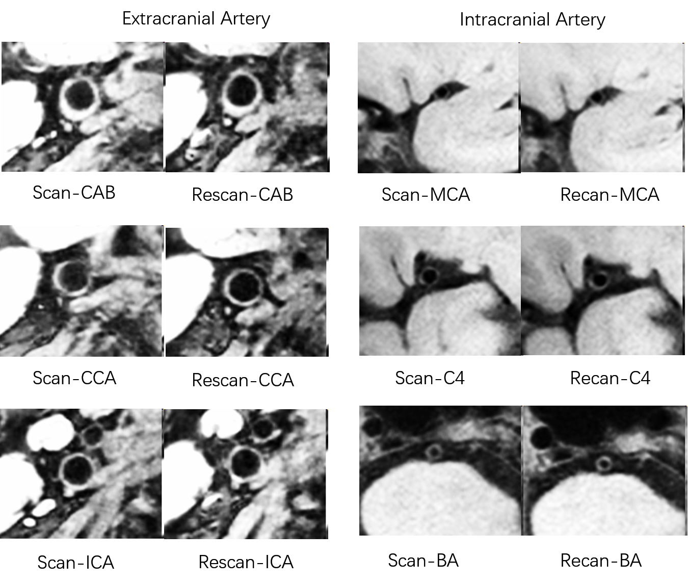
This study had ethical approval and informed consent was obtained from each volunteer and patient. 17 volunteers and 10 patients with ischemic stroke were scanned twice by the improved DANTE-SPACE sequence on a 3T MR system equipped with a 32-channel head and neck combined coil. Relevant imaging parameters were: spatial resolution 0.55×0.55×0.55 mm3, FOV 180 mm × 212 mm, number of slices 256, GRAPPA 2, turbo factor 48, scan time 9 min 6s. The time interval between the two scans were 1-2 weeks (mean 1.4 weeks) and 1–20 weeks (mean 10 weeks) for volunteers and patients, respectively. Five contiguous slices in the common carotid artery (CCA, 5 mm below the bifurcation), carotid artery bifurcate (CAB) and internal carotid artery (ICA, 5 mm above the bifurcation), as well as two reconstructed 2 mm thick slices in the distal basilar artery (BA), the distal intracranial carotid artery supaclinoid segment (C4), and the proximal middle cerebral artery (MCA) were used for morphological measurements. Lumen and vessel wall area and volume, mean and maximum wall thickness were measured for each scan. To test the scan–rescan reproducibility, all images were analyzed by one observer (one-year experience of vascular MRI). To test intra-observer reproducibility, the first scan was analyzed twice in a two-week separate by the same observer. The first scan was also analyzed by a second observer (six-year experience of vascular MRI) for inter-observer reproducibility.Results
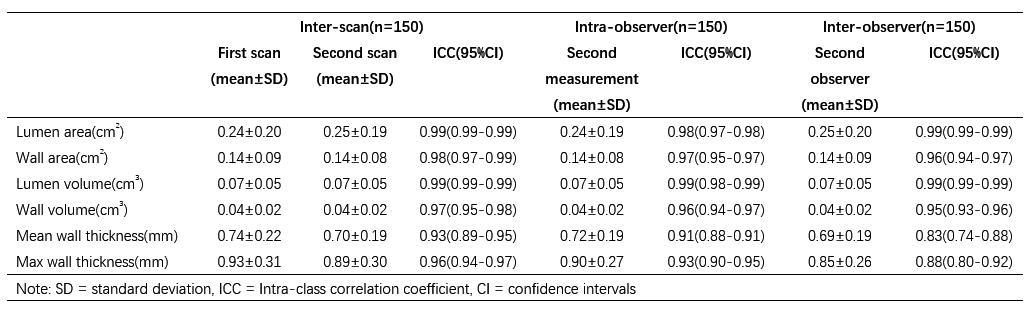
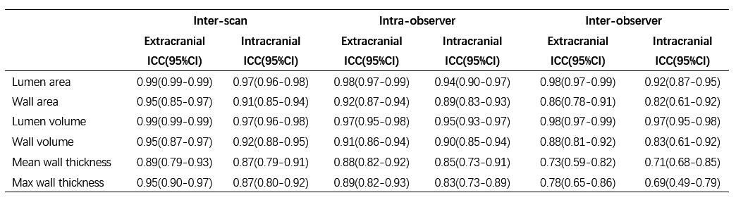
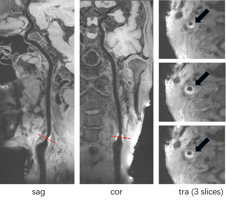
All 27 subjects completed the MR examinations. Two volunteers were excluded due to motion-induced poor image quality. The simultaneous intracranial and extracranial arterial vessel wall imaging method provided excellent delineation of both intracranial and extracranial arterial wall (Figure 1). All morphologic measurements, intra-class correlation coefficients (ICC) and 95% confidence interval were summarized in Table 1, which showed good to excellent reproducibility. For scan-rescan and intra-observer, the reproducibility was excellent with all ICCs were larger than 0.9. For inter-observer, the reproducibility was good for mean wall thickness (ICC=0.83) and maximum wall thickness (ICC=0.88), and excellent for other measurements with all ICCs larger than 0.9. Table 2 displayed the results of the reproducibility for patients and volunteers respectively. All ICCs of lumen and wall area and volume were above 0.9, while the ICCs of the mean and maximum wall thickness were range from 0.74 to 0.97. The ICCs of patients were generally higher than those of volunteers, especially for mean and maximum wall thickness. Table 3 summarized the reproducibility for extracranial arteries and intracranial arteries. The ICCs of extracranial arteries (range from 0.73-0.99) were higher than those of intracranial arteries (range from 0.69-0.97). For all analysis, inter-observer reproducibility was slightly lower but still good or excellent. Figure 2 showed representative vessel wall images of a patient with plaque in carotid bifurcation.Discussion and Conclusion
In general, this study demonstrated good to excellent reproducibility of scan-rescan, intra-, and inter-observer in one scan covered both intracranial and extracranial arteries, similar to previous studies on either intracranial or carotid arteries.3-7 The reproducibility of patients was higher than volunteers. This could be due to the thicker vessel wall thickness of patients than volunteers, which resulting a smaller error in the morphologic measurements. Meanwhile, the reproducibility for extracranial arteries was higher than intracranial arteries, which was benefited from the smaller partial volume effect of extracranial arteries. In conclusion, the improved 3D simultaneous vessel wall imaging technique is a reproducible MR method for intracranial and extracranial vessel wall imaging, which could potentially be used for monitoring therapy and disease progression in clinically.Acknowledgements
This work was supported in part by by National Natural Science Foundation of China (81327801), National Key R&D Program of China (2016YFC0100100), and Key Laboratory for Magnetic Resonance and Multimodality Imaging of Guangdong Province (2014B030301013).References
1. Xie Y, Yang Q, Xie G, Pang J, Fan Z, Li D. Improved black-blood imaging using DANTESPACE for simultaneous carotid and intracranial vessel wall evaluation. Magn Reson Med. 2016; 75: 2286–94.
2. Lei Zhang, Na Zhang, JunWu, Xin Liu, Yiu-Cho Chung. High resolution simultaneous imaging of intracranial and extracranial arterial wall with improved cerebrospinal fluid suppression. Magnetic Resonance Imaging. 2017;44: 65–71.
3. Feiyu Li, Vasily L. Yarnykh, Thomas S. Hatsukami, et al. Scan-Rescan Reproducibility of Carotid Atherosclerotic Plaque Morphology and Tissue Composition Measurements Using Multicontrast MRI at 3T. Journal of Magnetic Resonance Imaging. 2010; 31: 168–176.
4. E.S.J. Krönera,b, J.J. Westenbergc, R.J. van der Geestc, et al. High field carotid vessel wall imaging: A study on reproducibility. European Journal of Radiology. 2013; 82: 680– 685.
5. TWan-Qun Yanga, Biao Huanga, Xin-Tong Liub, et al. Reproducibility of high-resolution MRI for the middle cerebralartery plaque at 3 T. European Journal of Radiology. 2014; 83: 49–55.
6. Anouk L. M. Eikendal, Björn A. Blomberg, Cees Haaring, et al. 3D black blood VISTA vessel wall cardiovascular magnetic resonance of the thoracic aorta wall in young, healthy adults: reproducibility and implications for efficacy trial sample sizes: a cross-sectional study. Journal of Cardiovascular Magnetic Resonance. 2016; 18: 20.
7. Terry K. Koo, Mae Y. Li, BPS. A Guideline of Selecting and Reporting Intraclass Correlation Coefficients for Reliability Research. Journal of Chiropractic Medicine. 2016; 15: 155–163.
Figures