2998
The feasibility of a homemade dielectric pad using commercially available ultrasound gel with Gadolinium contrast material to improve B1 homogeneity for non-enhanced peripheral MR angiography1Tobata Kyouritsu Hospital, Kitakyusyu,Fukuoka, Japan, 2Tobata General Hospital, Kitakyusyu,Fukuoka, Japan, 3Radiology, UC, San Diego, La Jolla, CA, United States
Synopsis
We investigated an effect of homemade dielectric pads with using commercially available ultrasound (US) gel for improvement of B1 inhomogeneity in the peripheral artery examination using non-contrast fresh blood imaging (FBI) at 3T. We designed the two-bottle phantom mimics the iliac-femoral region, where often observed signal loss in peripheral non-contrast MRA due to B1 inhomogeneity. The result of the phantom study using US gel indicated uniform RF penetration in the B1 map. The US-gel pad improved the RF power penetration under the condition of B1 inhomogeneity and superior visualization of the left superficial femoral artery.
INTRODUCTION
The degradation of image quality due to B1 inhomogeneity is a cumbersome problem in the peripheral non-contrast fresh blood imaging (FBI) at 3T, which is often observed as a signal loss at the femoral region in thin patients.1 Dielectric pads have been reported to be useful to solve the B1 problem 2,3; however, commercially dielectric pads are not readily available, quite expensive, and limited in size and shape.4 Because ultrasound (US) gel have a dielectric property, 5,6 which can be readily available with less cost, we made “dielectric” pads with US gel with Gadolinium-based contrast agent (GBCA), and investigated the effect of the pads for improvement of B1 homogeneity at 3T.MATERIALS and METHODS
Commercially available US gel (SONO JELLY M, Toshiba Medical Supply Co., Japan) and Gadolinium-diethylenetriamine pentaacetic acid (Gd-DTPA; Magnevist®; Bayer Yakuhin, Ltd., Osaka, Japan) were used for dielectric pads. We made two types of US gel pad; US gel pad with GBCA (Gd-US-Gel pad) and without GBCA (US-Gel pad). The US gel itself has the high signal intensity in T2w images; the signals of US gel are reduced with increase in GBCA concentration. We set the optimal dilution to 4% GBCA. The US gel was placed into a plastic bag (20 x 15cm) and adjusted to a 2-cm thickness. In the phantom study, a femoral-mimicked-two-cylindrical phantom was prepared containing 0.32% (A) and 0.055% (B) copper sulfate solutions, which gave different RF penetration B1 values. The pads were placed on the surface of the femoral-mimicked-two-cylindrical phantom in 7 different patterns, as shown in Fig. 1. B1 measures were performed using a region of interest (ROI) and B1 value ratio index (BRI) on A and B in 7 patterns. The BRI difference between the A and B bottles was calculated as follows; BRI= {(B1 value of A) - (B1 value of B) / (B1 value of A)} x 100(%). Contrast ratio index (CRI) of femoral artery to vastus medialis muscle was calculated as follows; CRI(FA) = SI (right femoral artery) - SI(left femoral artery)/ SI (muscle). In addition, the BRI at the femoral artery in the volunteers was also measured. The effect of US-Gel and Gd-US-Gel pads was subjectively evaluated using a 4-point grading for improving the visualization of femoral arteries, (4; most clearly visualized, 3; more clearly, 2; fairly, and 1; poorly) by four radiation technologists. The paired t-test was used for all statistical analysis. All MR examinations were performed using a 3T clinical imager.RESULTS
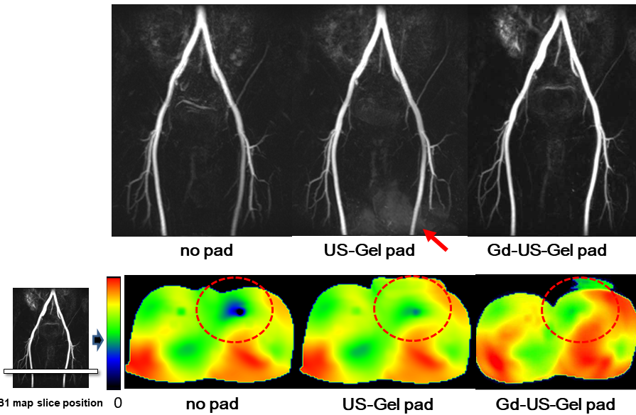
In the phantom study, patterns 4 and 5 placing the US-Gel pads on the B bottle shows lower BRI than patterns 2 and 3 placing on the A bottle (Fig. 1). Lower BRI indicates the improvement in B1 homogeneity (Fig. 2). Patterns 2 and 3 show higher BRI indicating inferior B1 homogeneity than the pattern 1 with no US-Gel pad. The effect of RF penetration was worse when placing the pad in between A and B bottles that is indicated by higher BRI in Fig. 2. In the volunteer study, average CRI(FA) significantly decreased with use of the pads (Fig. 3), indicating that the right and left femoral arteries are depicted similarly. There was no significant difference between US-Gel and Ga-US-Gel pads. The subjective evaluation of the femoral artery visualization improves with the US-Gel and Gd-US-Gel pads, as compared to no pad (Fig. 4). There was no significant difference in depiction of the femoral arteries using US-Gel and Gd-US-Gel pads. Figure 5 shows a typical example, with no pad, US-Gel pad, and Gd-US-Gel pad. The visualization of the left femoral artery indicate marked improvement with use of the pads. However, ambiguous high signal from the pad area overlapped obscures the image of the left femoral artery with US-Gel pad that is not seen with Gd-US-Gel pad.DISCUSSION
The homemade dielectric pad using US gel improves B1 homogeneity or RF penetration at the femoral region. However, image quality of FBI degraded by ambiguous signal artifacts using US-Gel pad, which might be caused by a slight movement as an imperfect subtraction between the systole and diastole and/or some fluctuations in TR intervals. Use of GBCA in the US gel sufficiently improves B1 homogeneity at the left femoral artery and suppress the high signal of US gel itself by shortening T1 of US gel that permits unambiguously visualization of both the left and right femoral arteries in FBI.CONCLUSION
A homemade dielectric pad with US gel and GBCA improves B1 homogeneity and evenly depiction of at the femoral arteries in FBI.Acknowledgements
No acknowledgement found.References
1) Yamamoto A, Nakamura K, et al. Non-contrast-enhanced MR Angiography of Peripheral Arteries at 3T MRI - Comparison with 1.5T -. Illinois Chicago, U.S.A. RSNA 2012.
2) Sreenivas M, Lowly M, et al. A simple solution for reducing artefacts due to conductive and dielectric effects in clinical magnetic resonance imaging at 3T. Radiology. 2007; 62(1):143-146.
3) Schmitt M, Feiweier T, et al. Improved uniformity of RF-distribution in clinical whole body-imaging at 3T by means of dielectric pads. ; The 12th ISMRM 2004
4) Marc D L, Daniel K, et al. High-Permittivity Thin Dielectric Padding Improves Fresh Blood Imaging of Femoral Arteries at 3T. Radiology. 2015; 50(2): 101-107.
5) Kataoka M, Isoda H, et al. MR imaging of the female pelvis at 3 Tesla: evaluation of image homogeneity using different dielectric pads. JMRI. 2007 Dec; 26(6): 1572-1577.
6) Kuroki Y, Yamamoto A, et al. The effect of a homemade dielectric pad with using commercially available ultrasound gel to correct B1 inhomogeneity in body MR angiography - a phantom study-. Saitama, Japan, The 44th JSMRM 2016.
Figures