2997
Free-breathing zoomed whole heart coronary MRA without respiratory gating using small-FOV 3D stack-of-stars radial sequence with pseudo-golden angle sampling1Radiology, Tokyo Metropolitan Police Hospital, Tokyo, Japan, 2Graduate school of Medicine, Division of Diagnostic Image Analysis, Tohoku University, Miyagi, Japan, 3MR Clinical Science, Philips Japan, Tokyo, Japan, 4Diagnosis of radiology, Tokyo Metropolitan Police Hospital, Tokyo, Japan
Synopsis
One of the problem of whole heart coronary MRA is the prolongation of acquisition time. It is caused for degrade image quality. However, the radial sampling technique is able to obtain image of inconspicuous artifact such as aliasing and motion; furthermore, the sequence is possible to reduce scan time by understate data sampling. Hence the zoomed whole heart coronary MRA with pseudo golden angle radial sampling was improved image quality without extend scan time.
Purpose
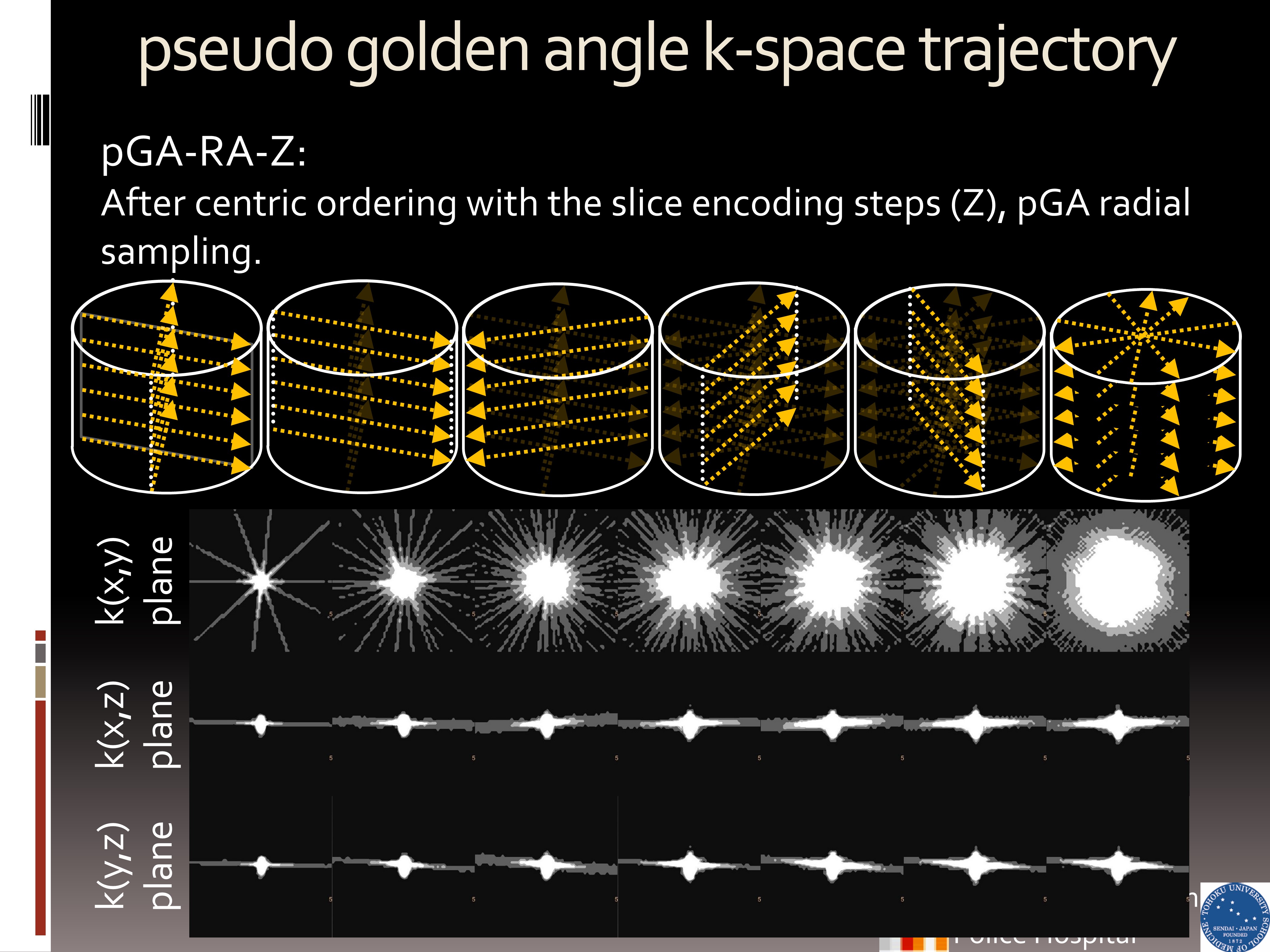
Whole heart coronary MRA (WHC-MRA), using balanced steady-state free-precession (bSSFP) with respiratory gating, is an established useful method for assessment of ischemic heart disease without contrast agents. One of the limitation of WHC-MRA is the acquisition time prolongation caused by the lower acceptance rate on navigator gating and irregularities in the breathing pattern, and this leads the deterioration of image quality. It is well known that the radial sampling sequence can significantly decrease a rigid motion artifact on liver and pelvic area. Moreover, radial scan can intrinsically reduce an aliasing artifact even if small FOV is applied. Recently, 3D stack-of-stars radial sequence with pseudo-golden angle radial sampling and the centric outer loop ordering (3D Vane) has been developed for free-breathing abdominal imaging. In this study, we demonstrate the feasibility of zoomed WHC-MRA without respiratory gating using 3D Vane combined with bSSFP on WHC-MRA.Methods and materials
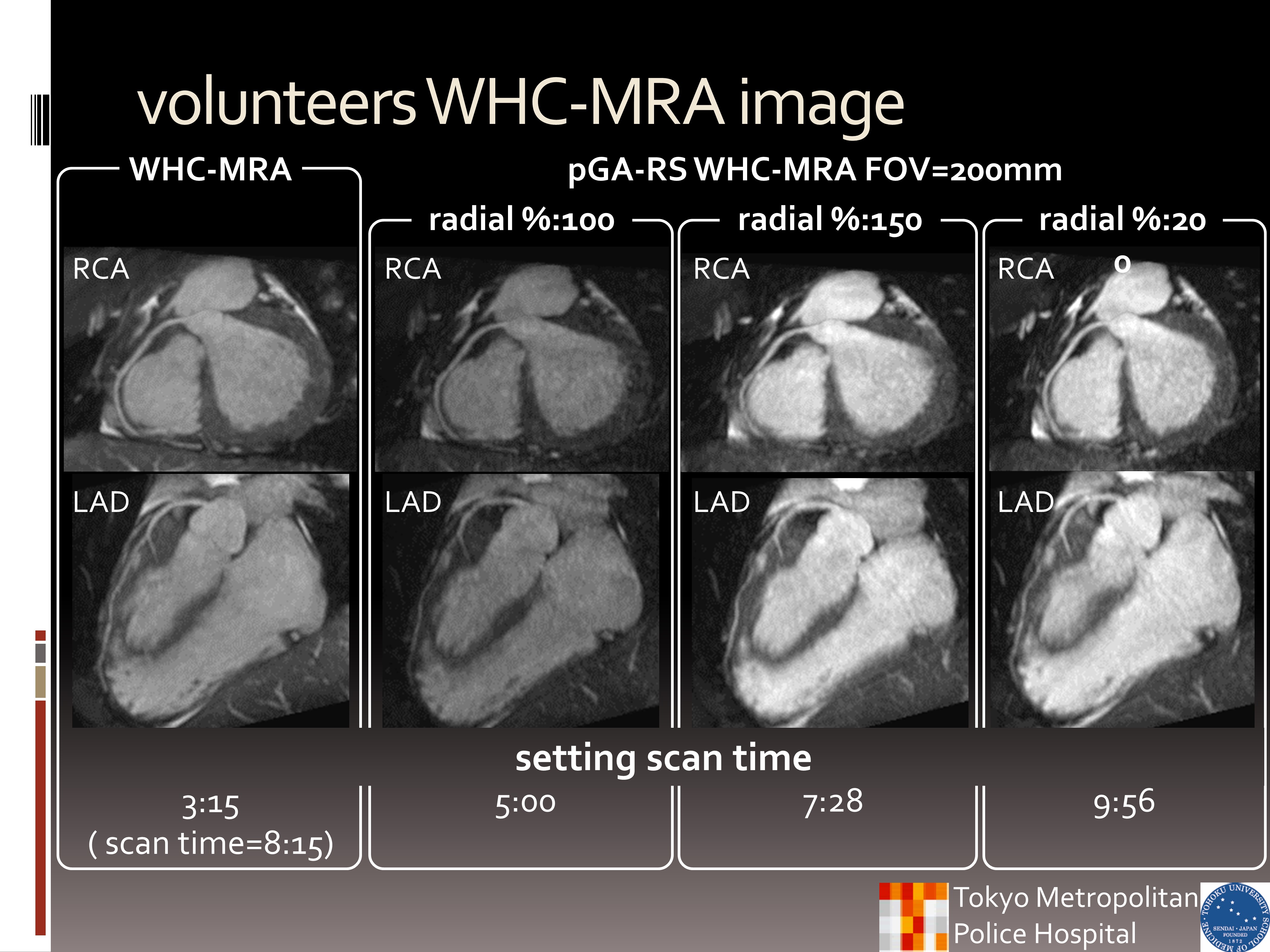
Using our institutional review board-approved procedures, 6 volunteers underwent WHC-MRA using a 1.5T Philips MR system and 32-channel torso-cardiac coil. WHC-MRA 3D bSSFP sequences were set to cover whole heart by axial slab orientation using navigator and vector cardiac gating with the following parameters: FOV (mm) = 350×315, in-plane resolution (mm) = 1.34×1.35, slice thickness (mm) = 1.7mm/0.85mm overlap, TR/TE (ms) = 3.4/1.72, SENSE factor = 1.5×2, NSA = 1, scan time= 3:04~4:05 (depend on heart beat and respiratory pattern). The parameters of zoomed WHC-MRA using 3D Vane without navigator gating were as follows: FOV (mm) = 200×200~350×350 (4 types of FOVs), radial%=100~200% (3 types of radial%s), in-plane resolution (mm) = 1.35×1.35, slice thickness (mm) = 1.7mm/0.85mm overlap, TR/TE (ms) = 3.8~4.1/1.88~2.1, SENSE factor = 2, NSA = 1, scan time= 5:00~9:56 (depend on heart beat). Images were assessed by using signal to noise ratio (SNR); contrast noise ratio (CNR), measured by left ventricle myocardium or blood signal intensity (SI) and standard deviation (SD); SNR efficiency (SNReff) and CNR efficiency (CNReff), calculated as the ratio of SNR or CNR to the square root of scan time. For the evaluation of aliasing, motion and streak artifacts, we evaluated using 5-point grades (grade “5” was absent, “1” was severe) by three blinded readers.Results
Coronary artery SNR of small FOV 3D Vane was significantly lower than that of conventional Cartesian 3D bSSFP sequence (P < 0.001). And increasing FOV and/or radial% improved SNR. CNR of 3D Vane was at least similar, or greater than that of 3D bSSFP sequence in some cases. Coronary artery SNReff and CNReff on small FOV 3D Vane were significantly higher than those of other sequences (P < 0.001). Motion and streak artifacts of 3D Vane were better than 3D bSSFP sequence (P < 0.001). Although 3D Vane was applied smaller FOV, it could effectively suppress aliasing artifacts.Discussion
The small FOV 3D Vane improved SNReff and CNReff compared to conventional sequences by decreasing scan time with the same in-plane resolution to 3D-bSSFP, because matrices can be reduced as well as FOV. 3D Vane could sufficiently suppress the motion artifacts without respiratory navigator gating by only an averaging effect of its central k-space of radial scan. In conclusion, zoomed WHC-MRA using 3D Vane can improve the image quality of WHC-MRA without scan time extension while preventing aliasing and motion artifacts.Acknowledgements
No acknowledgement found.References
Weber, Oliver M., Alastair J. Martin, and Charles B. Higgins. "Whole‐heart steady‐state free precession coronary artery magnetic resonance angiography." Magnetic resonance in medicine 50.6 (2003): 1223-1228.
Block, Kai Tobias, et al. "Towards routine clinical use of radial stack-of-stars 3D gradient-echo sequences for reducing motion sensitivity." Journal of the Korean Society of Magnetic Resonance in Medicine 18.2 (2014): 87-106.
Wundrak, Stefan, et al. "A small surrogate for the golden angle in time-resolved radial MRI based on generalized fibonacci sequences." IEEE transactions on medical imaging 34.6 (2015): 1262-1269. Zhou, Ziwu, et al. "Golden‐ratio rotated stack‐of‐stars acquisition for improved volumetric MRI." Magnetic Resonance in Medicine (2017).
Figures
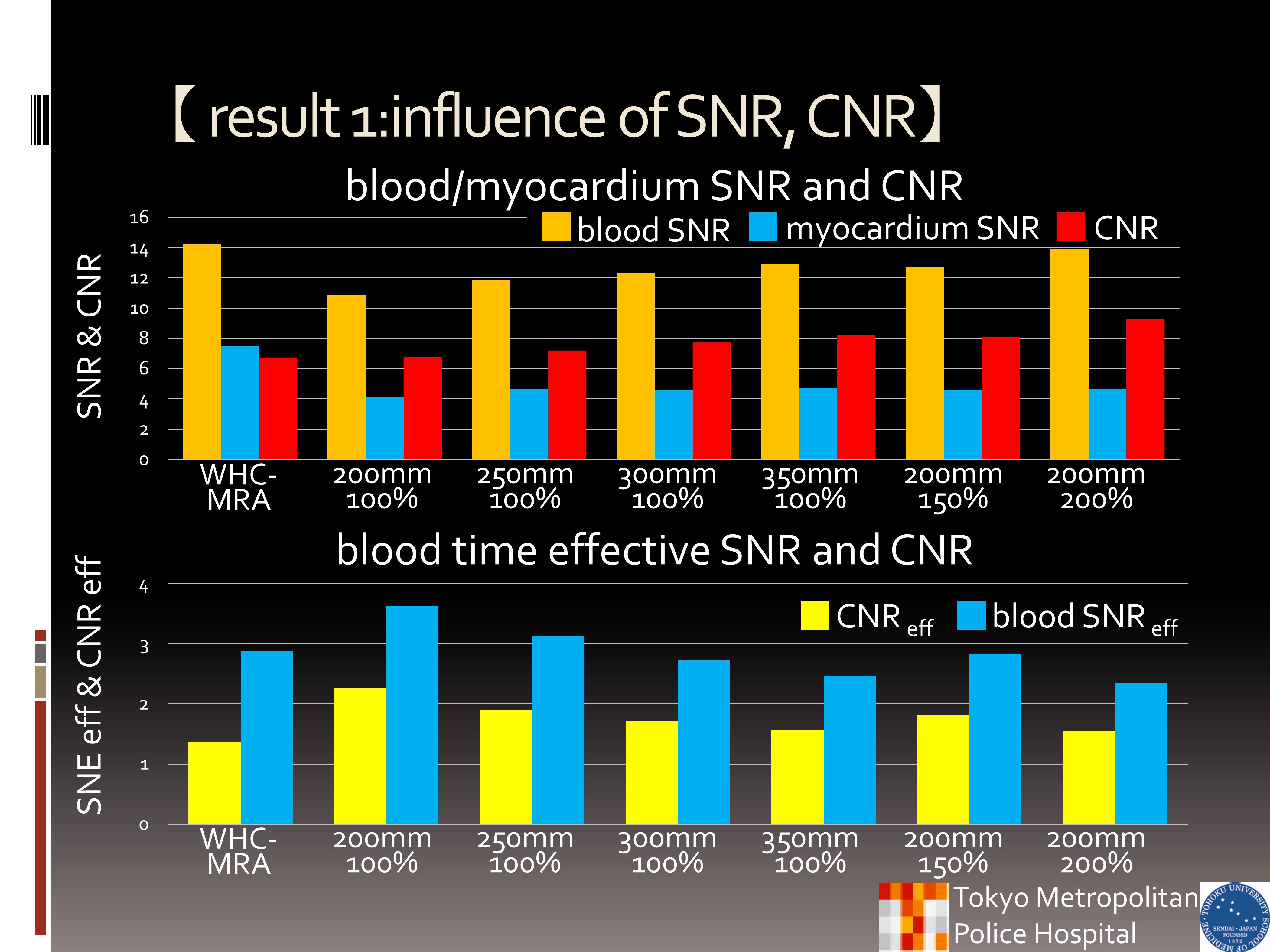
The SNR of blood was highest WHC-MRA. However, the CNR of blood/myocardium of all pGA-Rs WHC-MRA was higher than WHC-MRA. Moreover, the efficiency of SNR and CNR was higher than WHC-MRA.