2995
An accelerated peripheral MRA based on velocity-selective RF pulse using radial-MAGGULLI1Gachon University, Incheon, Republic of Korea, 2KAIST, Daejeon, Republic of Korea
Synopsis
We recently proposed a new peripheral MRA technique using velocity-selective gradient-echo (VS-GRE) sequence. Despite the high CNR and background suppression of the VS-GRE technique, this technique suffered from the reduced CNR efficiency, which was caused by the reduced sampling efficiency of radial trajectory in the peripheral region with anisotropic FOV. In this work, we propose a combination of the proposed peripheral MRA and the simultaneous multi-slice (SMS) imaging technique in the radial trajectory. In-vivo experiment results show that the proposed method could produce peripheral MRA with the reduced imaging time by radial-MAGGULLI.
Introduction
The SMS imaging techniques were developed to reduce imaging time without degradation of signal to noise ratio (SNR) by using the multi-band techniques. They were expanded to a non-Cartesian trajectory to utilize the benefits of the non-Cartesian trajectory and the SMS imaging techniques1,2, which used alternating RF phase for each radial spoke or view angle tilting gradient to increase the reconstruction efficiency of multi-slice images. Recently, we proposed a peripheral MRA technique using velocity-selective gradient-echo (VS-GRE) sequence3. Despite the high CNR and background suppression of the proposed method, this technique suffered from the reduced SNR efficiency, which was caused by the reduced sampling efficiency of radial trajectory in the peripheral region with anisotropic dimensions4. In this work, we propose a combination of the proposed peripheral MRA with the SMS imaging technique in the radial trajectory, which could increase the sampling efficiency of radial trajectory in an object with anisotropic dimensions1,2 using the inter-slice shifting in the radial trajectory2,5.Methods
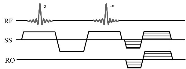
The sequence diagram of the proposed method is shown in Fig. 1. Multi-band RF pulse and inter-slice shift gradient are combined with VS-GRE sequence. The multi-slice image generation using intra-slice parallel imaging and inter-slice shifting (MAGGULLI)4 is extended to radial trajectory by modulating the magnitude of inter-slice shifting gradient (radial-MAGGULLI)2 according to the view angle as follows,
$$Gz=Acos(\theta+\phi)$$
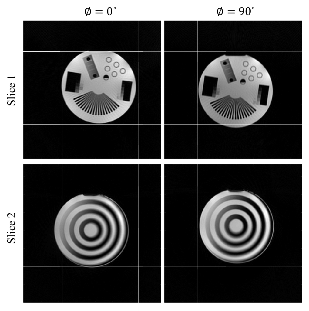
where Gz is the magnitude of inter-slice shifting gradient, A is the maximum magnitude of inter-slice shifting gradient, $$$\theta$$$ is the view angle of a radial spoke and $$$\phi$$$ is the offset of cosine modulation function. For the peripheral MRA, the offset of cosine modulation function ($$$\phi$$$) is selected to increase the efficiency of radial trajectory in the object with anisotropic dimensions (Fig. 2).
Results
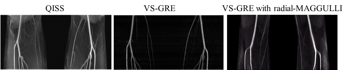
Phantom and in-vivo experiments were performed using a 3.0T MRI system (Siemens Magnetom Verio, Erlangen, Germany) with a peripheral angio matrix coil to verify the proposed technique. The MRA images were acquired using the following imaging parameters; field-of-view (FOV) = 380 × 380 mm2, slice thickness = 3 mm, flip angle = 40° and 30° for a single slice and SMS imaging, respectively, and TR/TE = 8.5/6 ms. In the proposed radial-MAGGULLI, we used SMS factor of two and view angle tilt = 30°. As shown in Fig. 3, the proposed method well reconstructed peripheral angiogram with the reduced acquisition time, where the imaging time of VS-GRE peripheral MRA without radial-MAGGULLI was 4m 15s and with radial-MAGGLLI was 2m 7.5s per a single station.Discussion & conclusion
In this work, we proposed an accelerated peripheral MRA based on the velocity-selective RF pulse using radial-MAGGULLI. A uniform spoke density of radial trajectory leads to a circular FOV, which reduces the sampling efficiency for the objects with anisotropic dimensions including a long peripheral region. In the proposed method, we can apply the inter-slice shift in any intra-slice directions by adjusting the offset of cosine modulation function ($$$\phi$$$) and can utilize empty regions of circular FOV in radial trajectory.Acknowledgements
This work was partly supported by the Basic Science Research Program through the National Research Foundation of Korea (NRF) funded by the Ministry of Education (NRF-2017R1D1A1B03033438) and a grant of the Korea Health Technology R&D Project through the KoreaHealth Industry Development Institute (KHIDI), funded by the Ministry of Health & Welfare, Republic of Korea (grantnumber : HI14C1135)References
1. Yutzy SR, Seiberlich N, Duerk JL, Griswold MA, Improvements in multislice parallel imaging using radial CAIPIRINHA. Magn. Reson. Med. 2011. 65(6).
2. Kim D, Kwon K, Kim B, et al., Simultaneous multi-slice (SMS) imaging technique for radial trajectory using inter-slice shifting gradient. ISMRM 2017.
3. Kim D, Seo H, Cho J, Kwon K, Han Y, Park H. Non-contrast-enhanced peripheral MR angiography using velocity-selective excitation. Magn Reson Med 2017.
4. Wu Z, Han F, Hu P. et al., Anisotropic field-of-view support for golden angle radial imaging. Magn Reson Med 2016. 76(1).
5. Kim D, Seo H, Oh C, et al., Multi-slice imAGe generation using intra-slice paraLLel imaging and Inter-slice shifting (MAGGULLI). Phy. Med. Bio. 2016. 61(4).
Figures