2992
A hybrid method combining Keyhole and segmented k-space filling for fast TOF imaging1Siemens Shenzhen Magnetic Resonance Ltd, Shen Zhen, China, 2Siemens Healthcare China Ltd, Shang Hai, China
Synopsis
In this work, we present a Keyhole method for fast Time Of Flight (TOF) imaging. We compare it with a recently published segmented k-space filling scheme. Moreover, we demonstrate the feasibility of combing the Keyhole and segmented methods for further acceleration. Such a hybrid TOF can be potentially suited for high-resolution angiograms at ultra-high field.
Introduction
Time Of Flight (TOF) MR angiography is a widely used MRI technique for visualizing brain arteries. It maximize the signals form the flowing tissue (arteries and veins) at the expense of the stationary tissue signal, where saturation bands are generally used to suppress the venous flow-related enhancement [1]. Various acceleration techniques have been proposed, including a recently published segmented k-spacing filling scheme [2]. In this study, we propose to apply the Keyhole [3] method to TOF sequence for scan acceleration. In vivo experiments were performed to investigate the feasibility of combining Keyhole and segmented TOF for an ultra-fast MRA scanning.
Method
The sequence diagrams and acquisition patterns of conventional TOF, segmented TOF and Keyhole TOF are shown on Fig1 and Fig2. In conventional TOF, venous saturation bands are used to eliminate possible venous enhancement effects [Fig1 a]. In Keyhole TOF, however, venous suppression bands are only applied when acquiring k-space center, since it is the k-space center that contains low spatial frequency information and thus determines overall image contrast, brightness, and general shapes. As shown in Fig2 [c1 and c2], in Keyhole TOF, the k-space is divided into two parts that acquired respectively with or without saturation pulse, the portion of which can be adjusted by a ‘keyhole factor’. In vivo experiments were performed on 4 health volunteers with a 3T Magnetom Spectra (Siemens Healthineers, 16 channels) with a16-channel head/neck coil. TOF parameters were: TE/TR = 3.74/23ms, flip angle=18, FOV = 200x180mm, slabs = 4, slices per slab = 40, slice oversampling = 20%, distance factor = -20%, base resolution = 384, voxel size = 0.3x0.3x0.5mm, slice partial Fourier = 7/8, acceleration in phase direction = 2, tracking band thickness/gap = 40/10mm, BW/pixel =186Hz. For comparison, 1) conventional TOF, 2) Keyhole TOF with keyhole factor = 50% and 33%, 3) segmented TOF with number of segment = 2 and 3, and 4) a combined Keyhole and segmented TOF were acquired.
Result
As shown in Fig3 [b and c], the Keyhole method accelerated TOF imaging as much as segmented k-space filling scheme. The former showed comparable image quality and better visibility of small artery braches then the latter [Fig3, green arrow]. Moreover, by combing the two methods, a scan time as fast as 3’27’’ can be achieved without sacrificing diagnostic information [Fig3 d and e]. It is also interesting to note that with segmented k-space filling scheme, the signals near scalp region are better suppressed than both Keyhole method and non-accelerated TOF [Fig2, yellow arrow]. The reason for this is still under investigation.
Conclusion
In this study, we demonstrate the feasibility of using Keyhole method for fast TOF imaging. It was shown to be comparable to the segmented k-space filling scheme regarding both time-saving and image quality. Moreover, a combination of the two methods has shown to decrease the scan time by 42% for further acceleration. Given that k-spacing filling scheme can reduce the short-term SAR [1], while with Keyhole method the usage of venous suppression bands is limited to k-space center acquisition, the combination of the two can offer further SAR reduction and thus be potentially suited for high-resolution angiograms at ultra-high field.
Acknowledgements
The author would like to thank Dr. Chen Shi (HKU) for her helpful suggestions and comments on this topic.
References
[1] Saloner D. An introduction to MR angiography. Radiographics 1995;15:453-465[2] Zhang Zihao Segmented TOF at 7 T MRI: Technique and clinical applications November 2015 Volume 33, Issue 9, Pages 1043–1050
[3] Joop J.van Vaals, et al, "Keyhole" Method for Accelerating Imaging of Contrast Agent Uptake
Figures
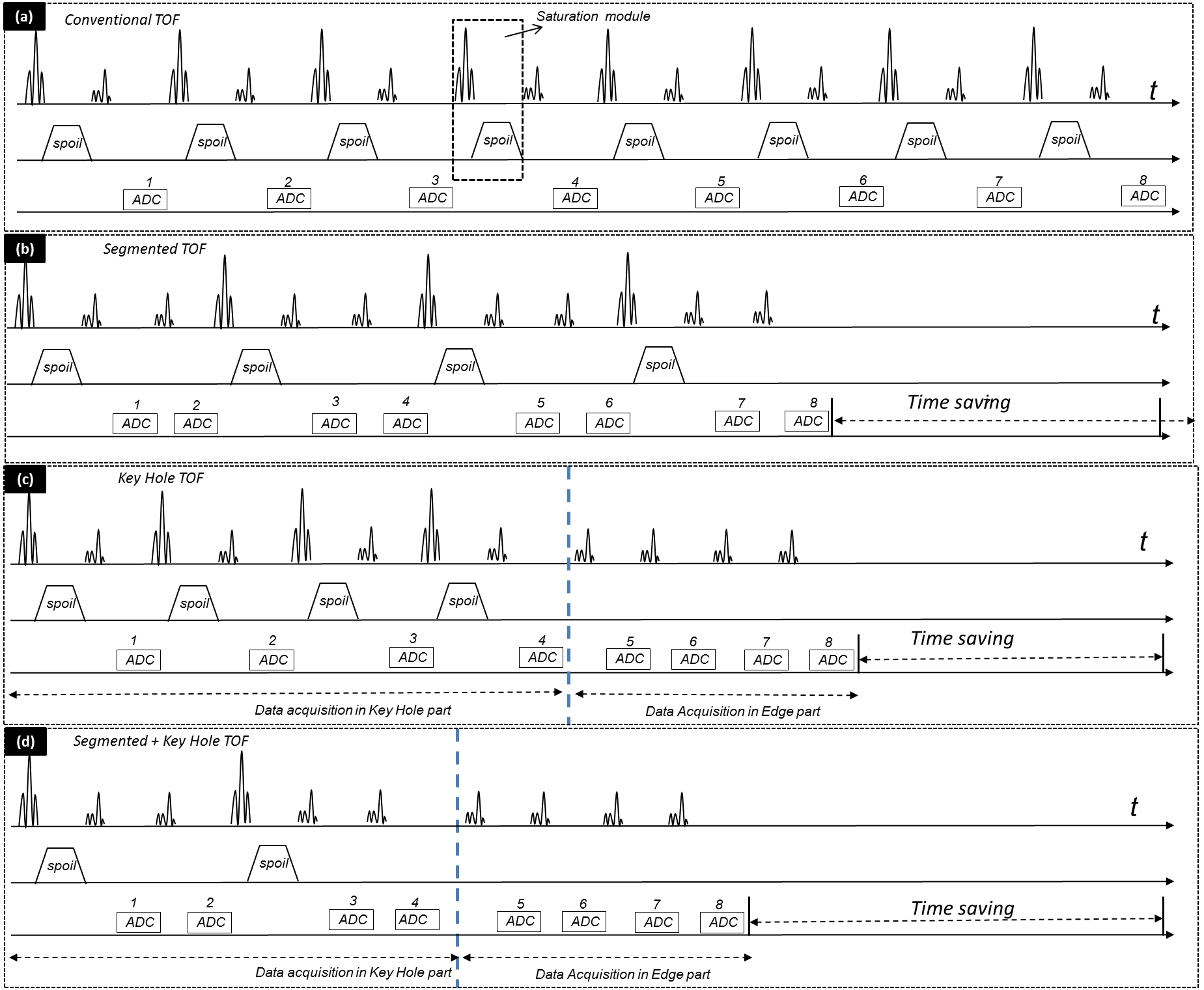
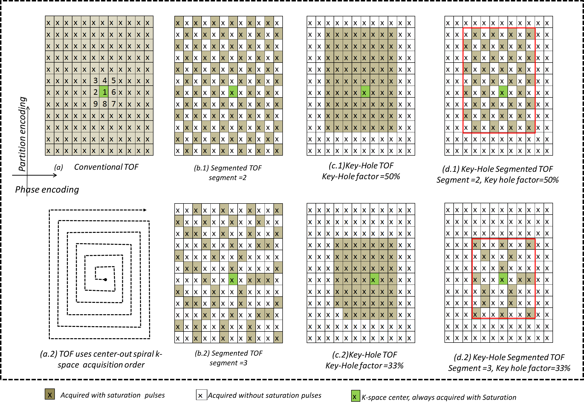
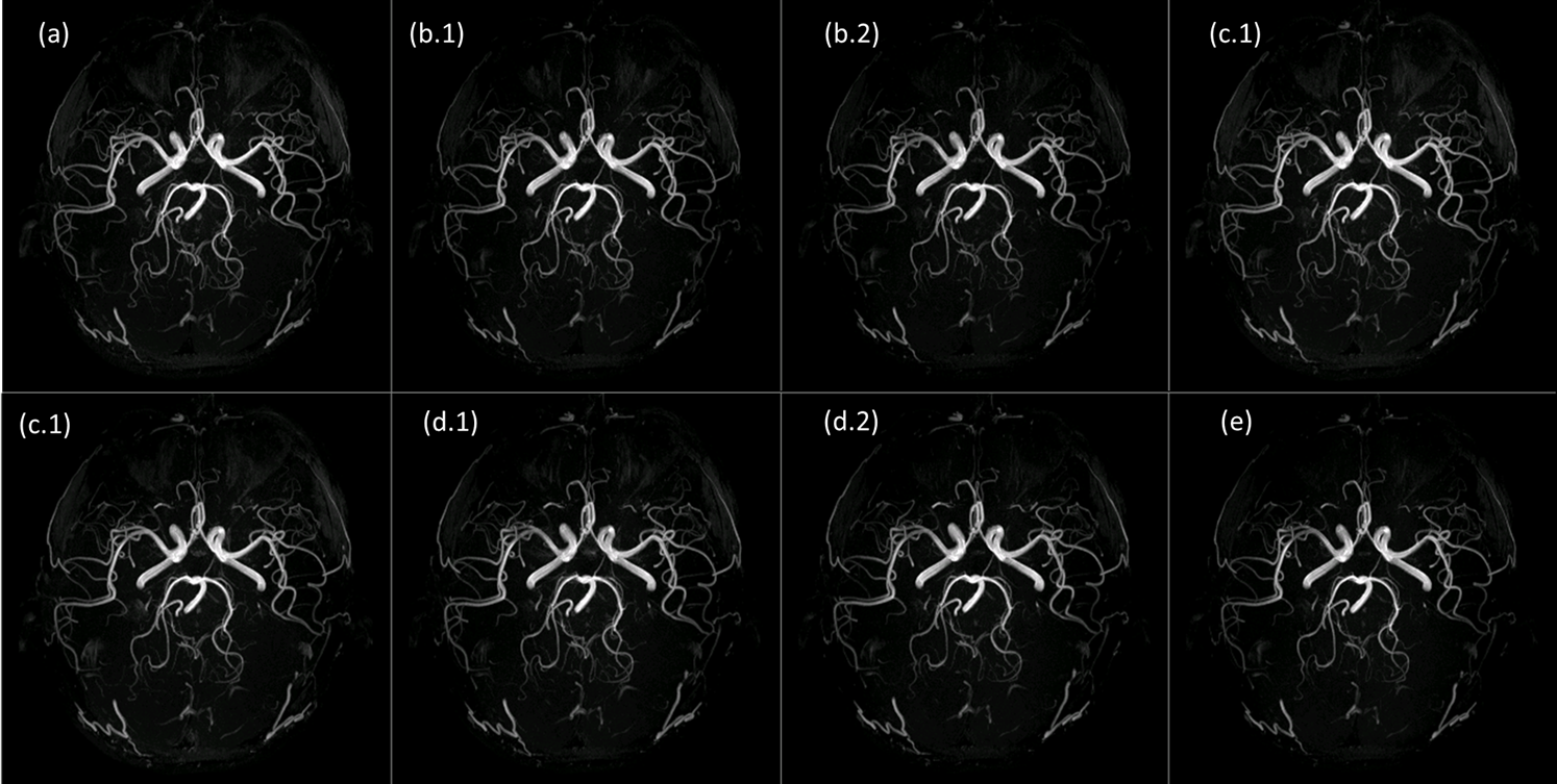
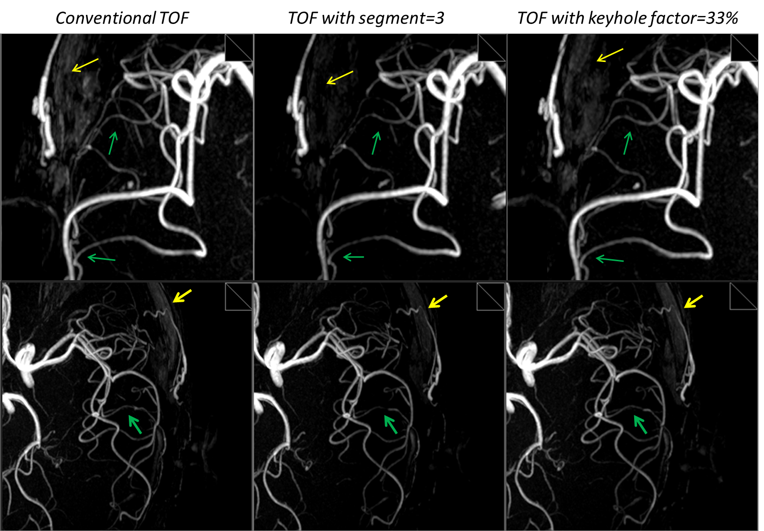
Fig4, comparable image quality and artery signals between different methods. Protocol parameters are the same as those for Fig3. From left to right: conventional TOF, scan time 5’59’’; segmented TOF, segment = 3, scan time 4’05’’; Keyhole TOF, keyhole factor 33%, scan time 4’05’’.