2982
Systematic evaluation of contrast-agent related image quality and vascular enhancement in abdominal time-resolved 4DMRA of minipigs1Radiology, University of Bonn, Bonn, Germany, 2MR and CT Contrast Media Research, Bayer AG, Berlin, Germany, 3Radiology, Neuroradiology, Sonography and Nuclear, Brüderkrankenhaus, Trier, Germany
Synopsis
This study systematically evaluated the impact of contrast agent (CA) doses both quantitatively and regarding image quality on time-resolved contrast enhanced MR-angiography (4D-MRA). The intra-individual study-design under highly standardized conditions was realized using an animal model. 5 anesthetized Göttingen minipigs received thoracic-abdominal 4D-MRA at 1.5T at five CA doses from 0.02-0.10 mmol/kgBW. We observed that the further the CA traveled along the circulation, the more a dose reduction resulted in weaker peak signal enhancement and low image quality. We conclude that CA dose reduction has varying effects on image quality in 4D-MRA with respect to vessel types and sizes.
Purpose
The passage of a contrast agent (CA) through the entire thoraco-abdominal vasculature can be visualized with highspatial and temporal resolution using time-resolved contrast enhanced MR-angiography (4D-MRA) techniques. Standard CA dosesthat have been proven effective for static MRA investigations are routinely used for 4D-MRA. However, several studies have notedhigh image quality of 4D-MRA also at reduced CA doses (1-5). However, systematic analyses of CA doses and resulting imagequalities of both arteries and veins were not yet performed and are crucial for identifying the optimum contrast protocol for evaluationof both arteries and veins (6). The purpose of this study was therefore to quantitatively and qualitatively evaluate the impact of CAdoses on 4D-MRA using an intra-individual study-design under highly standardized conditions in an animal modelMethods
Thoracic-abdominal 4D-MRA was performed on 5 anesthetized Göttingen minipigs using a standard whole-body 1.5Tscanner (Siemens Avanto Fit). Each animal received 4D-MRA at a standard dose (0.1 mL/kg body weight [=0.1 mmol/kg]) of 1Mgadobutrol (Gadovist, Bayer AG) and at reduced CA doses (0.08, 0.06, 0.04, 0.02 mmol/kg). CA were applied in a randomized ordereach with a flow rate of 2 ml/s and followed by a 20 ml saline chaser. Technical parameters of 4D-MRA were TR/TE/α =2.4ms/0.87ms/25°; TWIST (regions A/B, 20/25%); voxel size, 1.7x1.7x1.7mm³; image update time = 2s. Quantitative image analysiswas performed by ROI measurements in the pulmonary trunk, the abdominal aorta, the portal vein and the inferior vena cava.Qualitative image analysis was performed in 25 arterial and venous vessels by three independent radiologists.Results
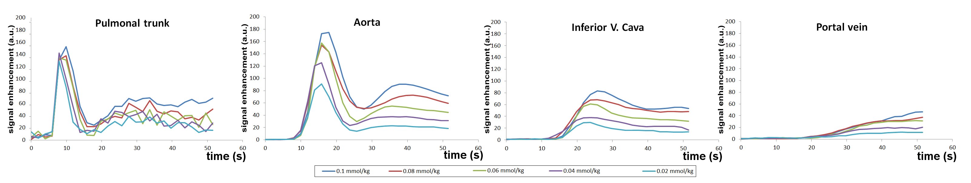
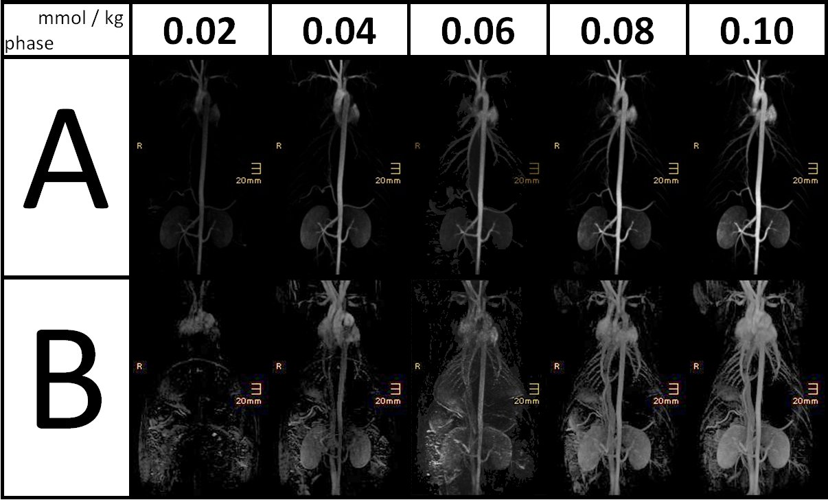
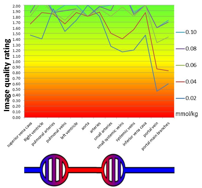
CA applications and 4D-MRA examinations were performed successfully in all animals and at all doses. Analysis of signalenhancement curves revealed shorter time-to-peak intervals and bolus durations with decreasing CA concentrations (fig.1). While atthe level of the pulmonary trunk the peak signal drop was little with lower CA doses, quantitative image analysis revealedsignificant linear decreases of peak signal intensities with dose reduction in the abdominal aorta (r²=0.96), the inferior vena cava(r²=0.99) and the portal vein (r²=0.99). However, while in arteries marked bolus peak signal intensities and thus high qualitymaximum intensity projections (MIP) still remained even at low CA doses, in veins peak signal intensities almost approached background noise levels at low CA doses resulting in low image quality of MIPs (fig. 2) and low image quality ratings (fig. 3).Conclusions
CA dose reduction in 4D-MRA results in narrower boluses and an almost linear decrease with dose in peak signal intensity afterpulmonary transit. Then, even at low doses steep bolus curves are observed in arterial vessels resulting in crispy vessel delineationshigh image quality ratings of arteries. After the next capillary transit peak signals substantially drop resulting in weak peaks invenous vessels at low doses and subsequently low image quality ratings of veins, particularly regarding the portal venous system.In conclusion, CA dose reduction has varying effects on vessel visualization in 4D-MRA with respect to vessel types and sizes withhigher doses being preferable to allow for complete 4D-MRA reports that include evaluation of both arteries and veins.Acknowledgements
No acknowledgement found.References
(1) Attenberger UI et al. Radiology 2010; 257: 879-887
(2) Morelli JN et al. Invest Radiol. 2012; 47(6): 376-82
(3) Kramer JH et al. Invest Radiol. 2013; 48(3): 121-8
(4) Krishnamurthy R et al. Radiographics 2016; 36: 523-537
(5) Hadizadeh DR et al. Proc.Intl.Soc.Mag.Reson.Med 2016; 24: 2679
(6) Hadizadeh DR et al. AJR 2012; 198: 1188-1195
Figures