2979
Pixel-wise quantitative myocardial perfusion mapping with cloud based non-linear iterative reconstruction using Gadgetron framework1NHLBI, NIH, Bethesda, MD, United States, 2University of Leeds, Leeds, United Kingdom, 3King’s College London, London, United Kingdom
Synopsis
In this abstract, we present a solution to speed up the non-linear reconstruction for myocardial perfusion imaging and demonstrate its clinical usage through the Gadgetron cloud deployed at Microsoft Azure infrastructure. We also achieved pixel-wise myocardial blood flow mapping on the non-linearly reconstructed images, given the computing power on the cloud. All these processing steps were inline integrated on the clinical MR scanners. As a result, the proposed solution allows us to deploy non-linear perfusion imaging with quantitative flow mapping as a clinical application.
Purpose
Myocardial perfusion imaging with saturation recovery has to trade SNR for spatial and/or temporal resolution required to meet the demands for stress imaging. Higher spatial resolution is desirable to minimize dark rim artifacts and to visualize thin walls [1]. To achieve higher spatial resolution while maintaining adequate temporal resolution, imaging at higher acceleration factors is required. Nonlinear reconstruction, such as L1 SPIRiT with motion correction (MOCO L1-SPIRIT) [2] or compressed sensing [3] can be used to improve image quality thereby making high acceleration factors possible. Incorporating MOCO into the reconstruction enables a free breathing stress perfusion study. Breath-holding has been a significant limitation of prior high resolution perfusion imaging methods using k-t approaches without MOCO. While these techniques significantly improve image quality, they have not been clinically feasible, mainly due to very high demand for computational power; that is, lengthy computation is a major limiting factor.
In this abstract, we present a solution to speed up the non-linear reconstruction for myocardial perfusion imaging and demonstrate its clinical usage through the Gadgetron cloud [4] deployed at Microsoft Azure infrastructure. We further utilized the computing power provided by the cloud to compute pixel-wise myocardial blood flow (MBF) maps from the non-linearly reconstructed perfusion image series. We showed these processing steps can be completed in ~3.5mins on the cloud. As a result, the proposed solution allows us to deploy non-linear perfusion imaging with quantitative flow mapping as a clinical application.
Methods
The MOCO L1-SPIRiT algorithm [1] was implemented in the Gadgetron framework [4]. This algorithm extends the L1-SPIRiT method by incorporating a motion correction operator which includes both forward and backward transformation to correct respiratory motion in perfusion image series. Unlike the original L1-SPIRiT, the unknowns in this algorithm are the motion corrected multi-channel complex images. A wavelet based L1-norm spatio-temporal regularization term was used to enforce signal consistency across different heart beats. Resulting perfusion image series were inputs for pixel-wise myocardial flow mapping. Intensities of every pixel were converted into the Gd concentration unit (mmol/L), using a Bloch-simulation method [5]. Gd signal of arterial input function and perfusion series were fed into MBF estimation module which has to process every pixel to get the MBF. All processing steps were integrated inline on the MR scanner using the Gadgetron framework. Resulting perfusion images and MBF maps were automatically computed and sent back to scanner. Prior in-line mapping protocols [5] used a matrix of 192×111 with 1.9x2.4 mm2 resolution.
To speed up the computation, iterative reconstruction and flow mapping were implemented in Gadgetron cloud. For the case of perfusion imaging, incoming k-space data for different slices is forwarded to different computing nodes, allowing constant computing time for any number of imaging slices. The cloud Gadgetron scheme was implemented in Microsoft Azure (https://azure.microsoft.com) and tested at King’s College London, UK (St Thomas’ Hospital). Typical cloud node has dual Intel Xeon E5-2673 v4 with 112GB RAM.
A free-breathing perfusion imaging protocol was tested: saturation recovery with b-SSFP readout, TR=2.7ms, FA 50o, FOV 360×270mm2, acquisition matrix size 256×144, 1.4x1.8 mm2 with 8 mm slice thickness, temporal interleaved acceleration R=4, PF=3/4, imaging duration 75ms, saturation time 105ms. N=5 patients underwent rest perfusion study with written consent. Gadolinium contrast agent (Gadovist, Bayer HealthCare, Germany) was administered as a bolus with a dose of 0.075 mmol/kg at 4 ml/sec with 20 ml saline flush. Raw k-space data were de-identified and converted to ISMRMRD format (no PII) and were sent to the cloud via an ssh connection. Images were returned to the scanner host where they are re-identified and inserted into the patient database in a seamless manner. All datasets were analyzed with both linear and non-linear reconstruction.
Results
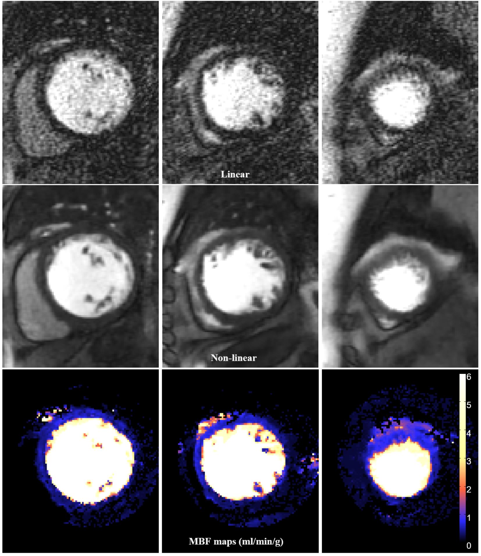
Fig. 1 gives example reconstruction and MBF maps.
The non-linear iterative method noticeably improved image quality and enabled
pixel-wise flow mapping at high spatial resolution. If a single computing node was
used, the imaging waiting time is 670s. With each slice being processed on its
own node, waiting time was reduced to 225s total after scan completion, for a
scan of 80 measurements. Conclusion
In
this abstract, we present a free-breathing, high resolution myocardial
perfusion mapping based on a cloud solution to speed up computation. High
spatial resolution and good image quality were achieved by using a non-linear
reconstruction. With inline integration on the MR scanner and the computational
power provided by cloud computing, these results demonstrate possible clinical
applicability of non-linear perfusion imaging.Acknowledgements
No acknowledgement found.References
[1] Plein S, et al., Eur Heart J. 29:2148-2155 (2008)
[2] Xue H, et al., ISMRM 1402 (2013)
[3] Otazo R, et al., MRM 64:767-776 (2010)
[4] Xue, H, et al., MRM 73:1015-1025 (2015)
[5] Kellman P, et al., JCMR 19:43 (2017)
Figures