2969
Right and left ventricular myocardial strain in healthy adolescents: Establishing normal reference values1University of Wisconsin - Madison, Madison, WI, United States
Synopsis
Tissue-tracking, a post-processing technique using routinely-acquired cine images, can assess strain, a multidimensional measure of myocardial contraction. In this prospective study, we measured left ventricular and right ventricular peak global radial, circumferential and longitudinal strain in 28 healthy adolescents ages 12-14 years. The data from this study provide normative global strain values to be used for future clinical and translational CMR studies.
Purpose
Early detection of abnormal cardiac function is critical in many adolescent cardiac diseases to enable prompt treatment prior to the onset of irreversible myocardial damage. Cardiac magnetic resonance imaging, with myocardial tagging,1 is considered the reference standard for quantifying left ventricular (LV) strain. However, this sequence requires a separate sequence for acquisition and is limited in its ability to assess right ventricular (RV) strain. Other techniques of assessing myocardial strain with CMR include displacement encoding (DENSE)2 and strain encoding (SENC)3 sequences, which also require a separate acquisition. More recently, methods have been developed to calculate LV and RV strain based on tracking tissue displacement on cine steady state free precession images, which are already part of all routine CMR studies. Although there is extensive data on its use in adults, there is still relatively little data on normal LV and RV strain values using tissue tracking (TT) in the pediatric population. The purpose of this study was to perform TT strain analysis in healthy adolescent volunteers to establish normal reference values for future clinical CMR and translational CMR research.Methods
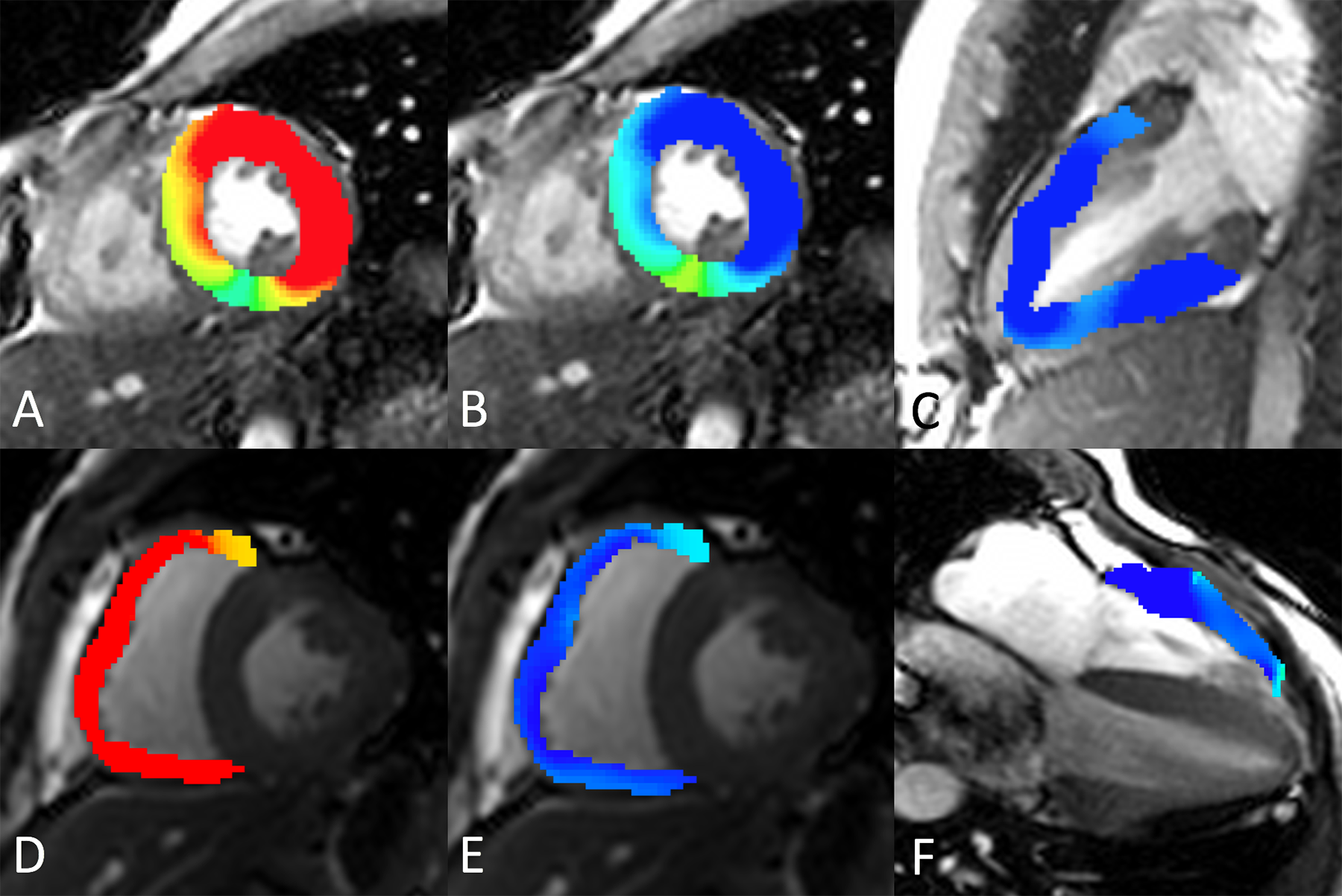
28 healthy adolescent volunteers (12-14 years, 9M/19F) were prospectively enrolled in this HIPAA compliant, IRB-approved study. CMR was performed on a clinical 3T scanner (MR750, GE Healthcare, Waukesha, WI). Short-axis and long-axis cine steady state free precession (bSSFP) series were acquired using a standard clinical protocol. CMR images were loaded into a commercially available CMR analysis software, cmr42 (Circle Cardiovascular Imaging, Inc., Calgary, Canada), for strain analysis using tissue tracking. LV and RV endocardial and epicardial contours were segmented at end-diastole on the short-axis and long-axis images. The LV and RV base and apex were also identified on the long axis views. These contours were propagated automatically to all phases of the cardiac cycle which, combined with tissue tracking, enable the assessment of LV and RV strain. Global radial, circumferential, and longitudinal strain values were recorded (Figure 1). Normative data for all volunteers and for M/F are reported as means ± standard deviation. Differences in strain values for M and F were assessed with the unpaired Student t-test.Results
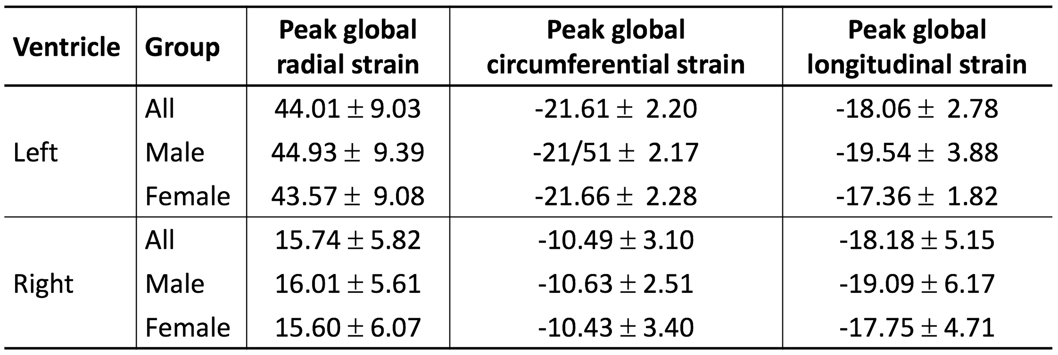
Normal global LV and RV strain values are summarized in the Table. Differences in strain values between M and F were not significant (p>0.05).Discussion
This study establishes normative LV and RV global strain values in 12-14 years old adolescents based on tissue tracking of cine bSSFP images. In contrast to what has been reported in adults, we observed no significant differences in global strain values between M and F in 12-14 years old healthy adolescents. The values reported here are higher, lower, and similar in magnitude for LV peak global radial, circumferential, and longitudinal strain assessed with similar techniques in adults.4 RV peak global circumferential and longitudinal strains we observed were slightly lower in magnitude than those reported in children and adults undergoing clinical CMR.5 These data will serve as a reference for future clinical and research CMR studies in patients with a variety of cardiac diseases.Acknowledgements
The Department of Radiology, University of Wisconsin - Madison receives research support from GE Healthcare.References
1. Axel L, and Dougherty L. MR imaging of motion with spatial modulation of magnetization. Radiology. 1989, 171: 841-5.
2. Kim D, et al. Myocardial tissue tracking with two-dimensional cine displacement-encoded MR imaging: development and initial evaluation. Radiology. 2004, 230: 862-71.
3. Osman NF, et al. Imaging longitudinal cardiac strain on short-axis images using strain-encoded MRI. Magn Reson Med. 2001, 46: 324-334.
4. Taylor RJ, et al. Myocardial strain measurement with feature-tracking cardiovascular magnetic resonance: normal values. Eur Heart J: Cardiovasc Imaging 2015; 16: 871–881.
5. Truong VT, et al. Cardiac magnetic resonance tissue tracking in right ventricle: Feasibility and normal values. Magn Reson Imaging 2017;38:189-195.
Figures