2948
Accelerating Dual Venc 4D Flow Using Compressed Sensing with Locally Low Rank along Velocity Encoding1Global MR Applications and Workflow, GE Healthcare, Menlo Park, CA, United States, 2Technical University of Munich, Munich, Germany, 3Radiology, Stanford University, Stanford, CA, United States, 4Electrical Engineering and Computer Science, University of California, Berkeley, CA, United States, 5Global MR Applications and Workflow, GE Healthcare, Munich, Germany
Synopsis
Dual Venc has been developed to improve the accuracy of conventional 4D flow in high dynamic range of velocity. However, dual Venc acquisition doubles scan time. This work explored a high dimensional compressed sensing method to accelerate dual Venc 4D flow by utilizing additional data redundancy in the velocity encoding dimension.
Introduction
Recent years, 4D flow has demonstrated great potential to resolve volumetric flow in all directions and thus enables comprehensive assessment of hemodynamics-related pathologies. While 4D flow attempts to resolve velocity in a large volume and all cardiac phases with a high dynamic range, conventional 4D flow requires selection of a single velocity encoding (Venc) higher than the peak flow to avoid phase wrapping, which compromises accuracy in regions and at cardiac phases with relative low velocities. A dual Venc technique was developed to address this limit by collecting high and low Venc images [1]. However, the two Venc acquisition significantly increases scan time. In this work, we aimed at accelerating dual Venc 4D flow imaging by exploiting sparsity in the velocity encoding dimension with compressed sensing (CS) reconstruction.Methods
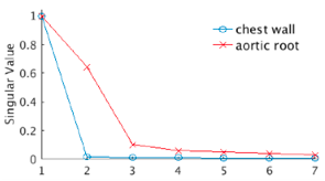
Typically, dual Venc 4D flow encodes 3D (x, y, z) velocity in two Venc’s using a 7-echo acquisition scheme. To put it simply, the signal at a voxel in an echo image can be expressed as M · exp(i·(Δφ + π(cxvx + cyvy + czvz)/Venc)), where Δφ indicates background phase offset (e.g. eddy current effects, Maxwell term, etc), vx, vy and vz are the 3D flow velocities and cx, cy and cz are constants representing the velocity encoding scheme of the specific phase encoding echo. Clearly, signal in the 7 echoes manifests high linear correlation. Furthermore, background phase varies very slowly in space and flow field also exhibits certain spatial continuity in most regions, indicating low rank in a data matrix comprised of 7 echo signals (columns) in a local spatial neighborhood (rows). Figure 1 shows the sharp decrease of the SVD single value at a background region in chest wall and a fast flow region in aortic root. Such data redundancy could be utilized to improve reconstruction from sparse data.
To evaluate the proposed method, whole-heart 4D flow was performed in 3 healthy volunteers on a GE 3T 750 system. A dual Venc sequence was scanned with low Venc encoding of 80 cm/sec followed by high Venc encoding of 200 cm/sec in each segmented data acquisition. Full k-space data was collected and retrospectively undersampled to simulate different accelerations and reconstructions. For high-dimensional CS, a variable density Poisson disk sampling was used with the sampling pattern shifting along both cardiac phases and velocity encoding echoes. In reconstruction, first, sensitivity maps were estimated from fully sampled central k-space data using ESPIRiT [2]. Then, each dataset was processed using 3 reconstructions in a l1-ESPIRiT framework exploring: 1. CS in space: spatial sparsity using wavelet in [y, z], 2. CS in space & time: spatial sparsity as in 1 and additional temporal sparsity along cardiac phases using total variation (TV) and 3. CS in space, time & echo: spatial and temporal sparsity as in 2 and additional data sparsity in the velocity encoding dimension using locally low rank (LLR) performed separately at each cardiac phase. At last, dual Venc combination was conducted after background phase correction and the high velocity to noise (VNR) image with low Venc was unwrapped based on the unaliased high Venc image.
Results
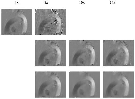
Figure 2 compares superior-inferior flow at a systolic phase obtained using different reconstruction methods and accelerations. CS based on spatial sparsity only produces large artifacts at 8x. Adding temporal TV substantially suppresses the aliasing artifacts. However, its flow image suffers from increasing artifacts and noise with higher acceleration. In comparison, from the same datasets, the proposed reconstruction with LLR along velocity encoding provides more accurate flow that is similar to the full acquisition and enables acceleration higher than 10x.Discussion
This worked demonstrated a new reconstruction that fully explores sparsity in dual Venc 4D flow along all dimensions. Our preliminary results show that the data redundancy along velocity encoding can be exploited to improve reconstruction and enable highly accelerated dual Venc 4D flow imaging.Acknowledgements
No acknowledgement found.References
1. Callaghan FM, MRM 2016. 2. Uecker M, MRM 2014.Figures