2938
Blood flow measurement using 3D cine PC MRI within the abdominal aortic aneurysm and visceral arteries in pre- and post-EVAR condition; blood flow in the SMA might be improved after EVAR.1Radiology, Hamamatsu University School of Medicine, Hamamatsu, Japan, 2Department of Fundamental Development for Advanced Low Invasive Diagnostic Imaging, Nagoya University, Graduate School of Medicine, Nagoya, Japan, 3Applied Science Laboratory Asia Pacific, GE Healthcare Japan, Hino, Japan, 4Radiology, Stanford University School of Medicine, Stanford, CA, United States, 5Radiology, Nagoya University School of Medicine, Nagoya, Japan
Synopsis
The blood flow volume within the visceral arteries were measured and compared between pre-and post-EVAR conditions using 4DFlow MRI. The maximum systolic flow volume ratio in the SMA to that of the aorta showed significant increase after EVAR. 4DFlow might be useful for evaluation of the blood flow dynamics of the aorta and visceral arteries in pre- and post-EVAR condition.
Introduction
Abdominal aortic aneurysm (AAA) involves significant deformity of the aorta. In such vessels, abnormal blood flow dynamics is more likely to appear. The fluctuated blood flow dynamics may spoil the kinetic energy of the blood flow(1), and subsequently blood flow delivery might be less efficient.
Endovascular aneurysm repair (EVAR) is widely performed to prevent AAA from rupture. Besides the primary aim, EVAR may also contribute to the repair of deformed blood pathway and provide a proper blood flow passage. This may reduce the turbulence of blood flow, relieve the loss of blood flow kinetic energy, and subsequently provide more efficient blood delivery to the visceral arteries.
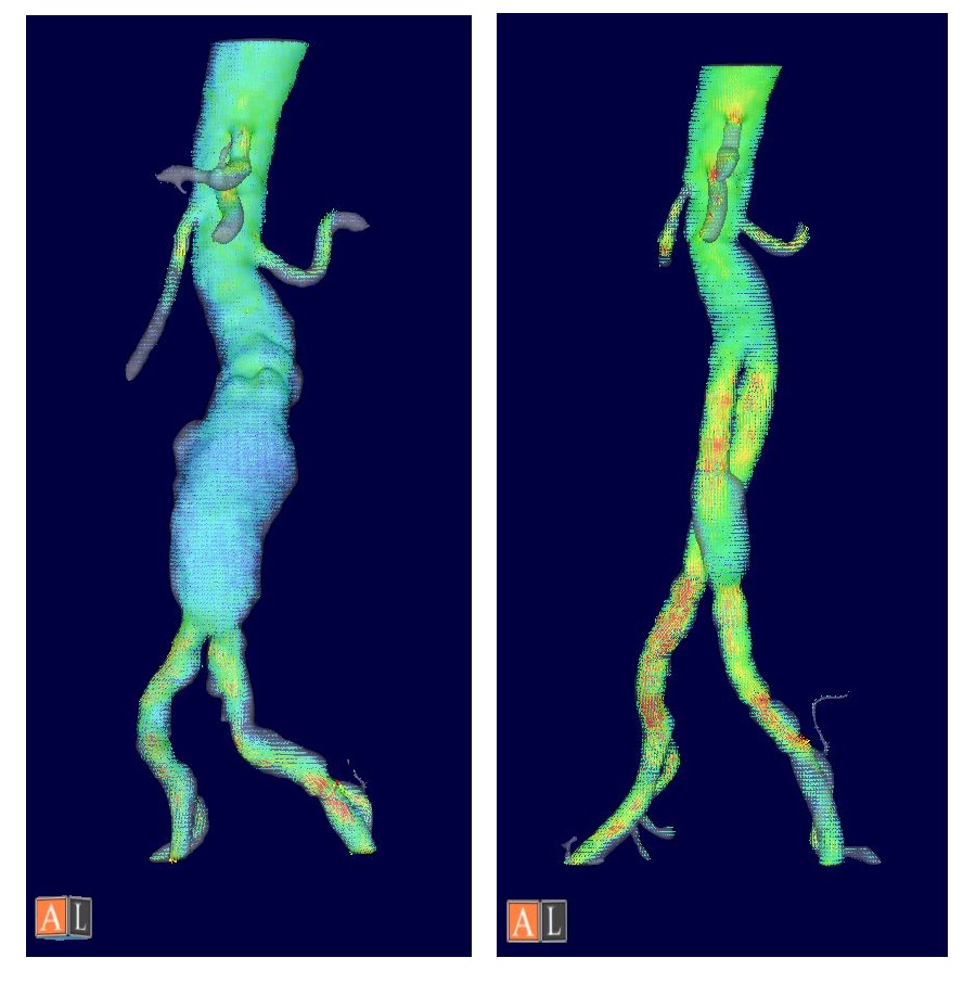
Recently developed 3-Dimensional cine phase contrast MR imaging (4DFlow) has enabled the coverage of full spatial and temporal data of the velocity vectors of the flowing blood within the whole abdominal aorta, thereby allow blood flow dynamics analysis with an aid of flow analysis software (figure 1).
In the present study, we aimed to evaluate the effect of EVAR treatment to the blood flow profiles of visceral arteries, by comparing the blood flow within the visceral arteries between pre-and post-EVAR conditions using 4DFlow MRI.
Materials and methods
Nine patients (67 to 80 y.o.) with AAA underwent MRI scan including Cartesian 4DFlow between September 2013 and January 2015. All patients had infrarenal fusiform aneurysm and were treated by EVAR.
The MRI were performed for all patients within 1 year before and after EVAR operation. All the MR studies were conducted on 3.0T MR imagers with phased array coil (Discovery 750 and Discovery 750W, GE Healthcare, Waukesha, WI). ECG gated, respiratory compensated gradient-echo-based coronal 4D-Flow covering from suprarenal abdominal aorta to common iliac arteries was performed following the contrast enhanced 3D MR Angiography (Gd-3DMRA) for the determination of the aortic boundary.
The parameters set for 4DFlow data acquisition are as follows; TR/TE/FA/NEX of 4.5-5.5/2.0-3.0/15/1, FOV of 32 cm, Matrix of 224x224, 2 mm thickness, 60 partitions, 20 /cardiac cycle.
2D cine PC with velocity encoding (VENC) of 200 cm/s was performed placing a transverse section within thoracic aorta prior to 4D-Flow acquisition. The VENC for 4DFlow acquisition was set to the max flow velocity of 2DcinePC + 20cm/sec.
The acquired 4DFlow data was postprocessed using an flow analysis software (Flova II, R’tech, Hamamatsu, Japan). The blood flow vector data derived from 4DFlow and the geometric boundary of the aorta determined by Gd-3DMRA were interpolated.
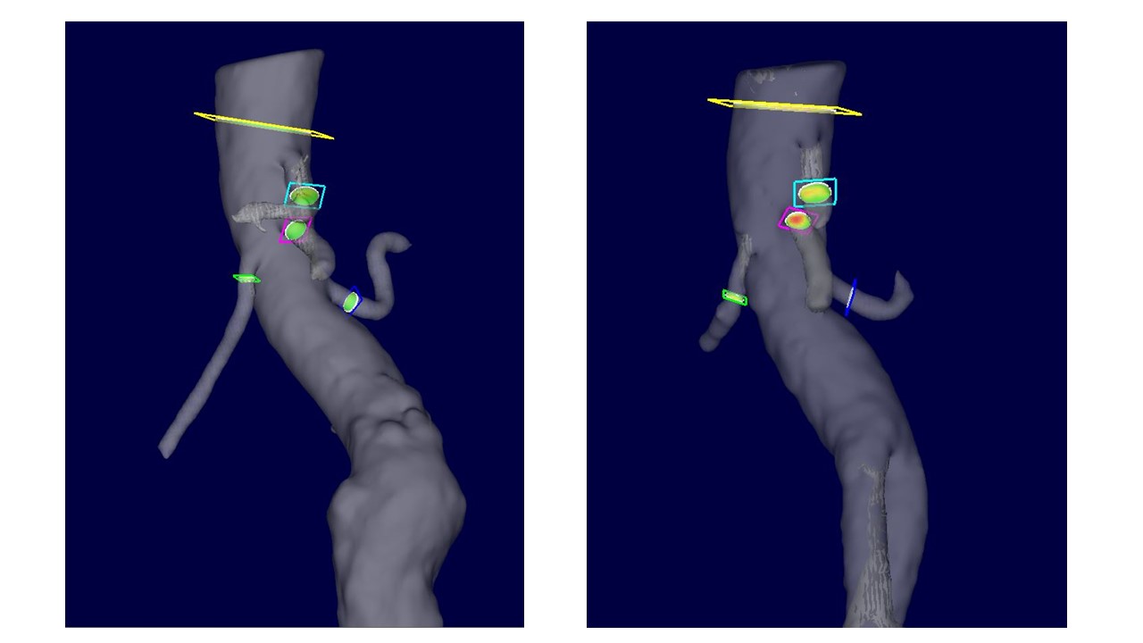
The flow volume at the sections in suprarenal aorta (SupraRA), celiac artery (CA), superior mesenteric artery (SMA) and renal arteries (RA) were measured (figure 2). Each datasets consisted of flow volume data of 20 phases per cardiac cycle. The flow volume ratio (FV-ratio) of the visceral arteries compared to that of SupraRA were calculated. Time-averaged, maximum systolic, average systolic (1-7th phase), early diastolic (8-12th phase), late diastolic (13-20th phase) FV-ratio between pre- and post-EVAR condition were statistically analyzed using non-parametric test (Wilcoxon’s signed-rank test, p≤0.05 were considered to be significant).
Results
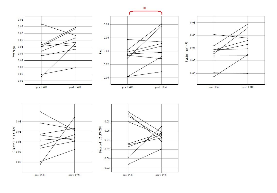
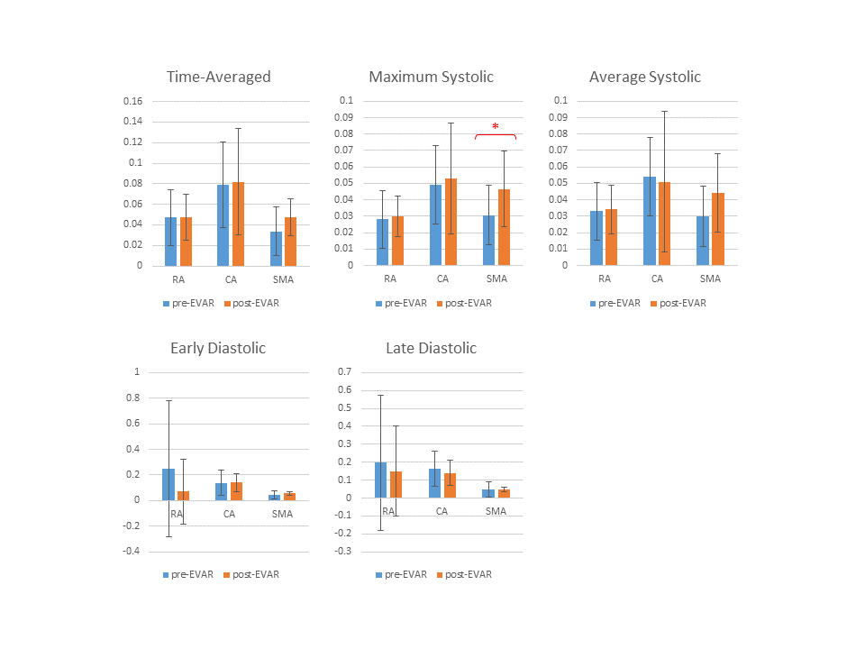
Seven CAs, nine SMAs and eighteen RAs were measured in nine patients. CA could not be segmented in two cases by insufficient contrast in MRA.
The time-averaged FV-ratio within the measured visceral arteries showed no significant change between pre- and post-EVAR. The maximum systolic FV-ratio has significantly increased in SMA after EVAR treatment (p<0.05). No significant change in FV-ratio was observed in SMA in other cardiac phases. No significant change in FV-ratio were observed within CA, RAs throughout all cardiac phases.
Discussion
EVAR repairs deformed blood pathways, which may be contributing to improved blood flow passage within the aorta. The significant increase of the maximum FV-ratio within the SMA after EVAR treatment was observed in this study.
The total FV-ratio of the visceral arteries showed no significant change between pre- and post-EVAR. This was understandable since the demand for blood in each vessels should not change after EVAR. All measurements were performed at fasting state skipping one meal before the MR scan.
Interests have not been paid concerning the hemodynamic changes of the visceral arteries after EVAR. Current study has shown that the placement of endovascular stent affects visceral hemodynamics even without the occlusion of the orifice.
Conclusion
4DFlow measurement showed the possible improvement of blood flow delivery in the SMA after EVAR. 4DFlow might be useful for evaluation of the blood flow dynamics in pre- and post-EVAR condition, not only in the aorta but in the visceral arteries.Acknowledgements
No acknowledgement found.References
(1) Burris, N. S. and M. D. Hope (2015). "4D flow MRI applications for aortic disease." Magn Reson Imaging Clin N Am 23(1): 15-23Figures