2931
Flow-encoding Arterial Structure Acquired using Silent-MRA: A Preliminary Study1Department of Radiology, Wan Fang Hospital, Taipei Medical University, Taipei, Taiwan, 2GE Healthcare, Taipei, Taiwan, 3Department of Radiology, School of Medicine, College of Medicine, Taipei Medical University, Taipei, Taiwan
Synopsis
Silent magnetic resonance angiography (Silent-MRA), which combines the Silent Scan algorithm to achieve a zero echo time with an arterial spin-labelling method, has recently been introduced as a novel MRA technique. Many studies of Silent-MRA focused on evaluating vascular structure; however, reports of further investigations into the flow information generated by Silent-MRA cannot be found. To this end, we compared the flow-encoding Silent-MRA signal with phase contrast flow imaging and found a linear correlation between the two. This preliminary study demonstrates the potential power of using flow-encoding Silent-MRA in assessing complicated vascular disease.
INTRODUCTION
Silent magnetic resonance angiography (Silent-MRA) is a novel imaging technique that integrates a continuous arterial spin-labelling (ASL) strategy and a three-dimensional (3D) radial sampling to achieve a nominal echo time of zero (ZTE). Previous reports have shown its ability to assess arterial structures in blood vessels with endovascular therapeutic devices such as intracranial stents [1] and in coiled intracranial aneurysms [2], which cannot be visualized using a traditional time-of-flight (TOF)-MRA. This is because Silent-MRA utilizes ZTE inside-out radial acquisition by minimizing the phase dispersion of the labelled blood flow signal and decreasing magnetic susceptibility [1]. Many studies using Silent-MRA focused on evaluating vascular structure, and reports of further investigations into the flow information generated by Silent-MRA are nonexistent. The aim of this study was to understand whether the flow information obtained by Silent-MRA is reliable by comparing the flow-encoding Silent-MRA signal distribution with phase contrast flow imaging.
METHODS
One male participant (26 years-old) jointed in this study. All MRI acquisitions were performed using a 3T clinical scanner (Discovery MR750w; GE Healthcare, Milwaukee, USA) using a geometry-embracing method (GEM) head-and-neck unit for signal detection and a whole-body coil for radio-frequency excitation. Both the Silent-MRA and TOF-MRA scans were applied to acquire the arterial structure. The Silent-MRA was performed using these parameters: TR, 1116.4 ms; flip angle, 5°; FOV, 18 cm; matrix, 256 × 256; section thickness, 1.2 mm; NEX, 1.5; bandwidth, 20 kHz; and acquisition time, 7 minutes 40 seconds. The TOF-MRA was performed using these parameters: TR/TE, 26/3.4 ms; flip angle, 15°; FOV, 22 cm; matrix, 512 × 384; section thickness, 1.6 mm; NEX, 1; bandwidth, 41.7 kHz; and acquisition time, 3 minutes 31 seconds. In the flow-measuring scan, the Non-Invasive Optimal Vessel Analysis (NOVA; VasSol, Inc., Chicago, USA) system was used to perform quantitative flow measurements in the following 7 cerebral arteries: the basilar artery (BA), the left and right anterior cerebral arteries (ACAs), the left and right inferior cerebral arteries (ICAs), and the left and right middle cerebral arteries (MCAs).
RESULTS and DISCUSSIONS
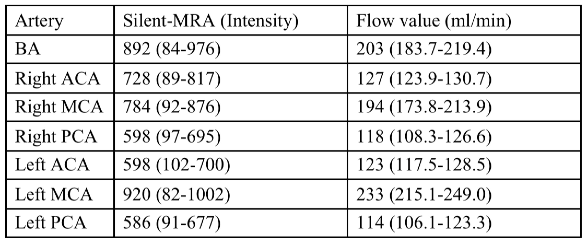
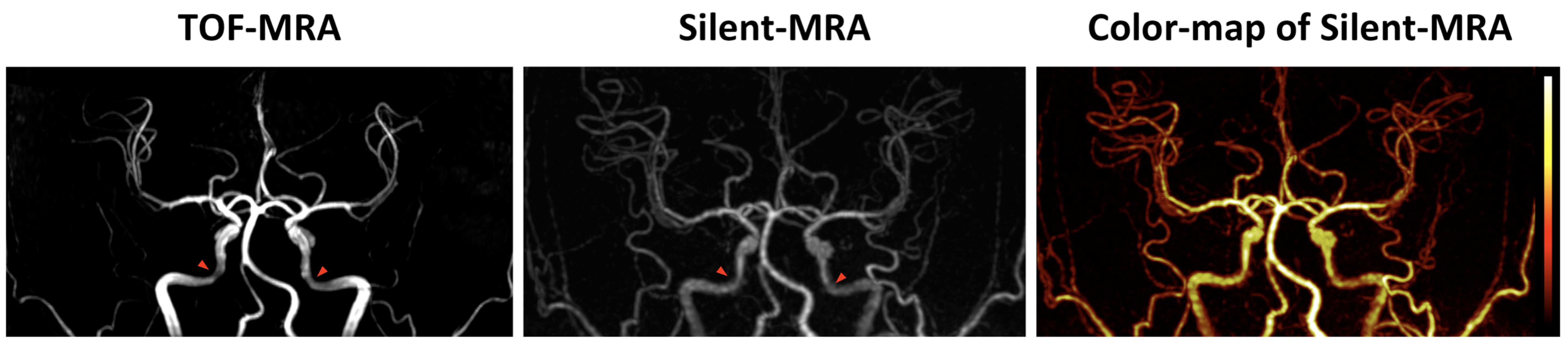
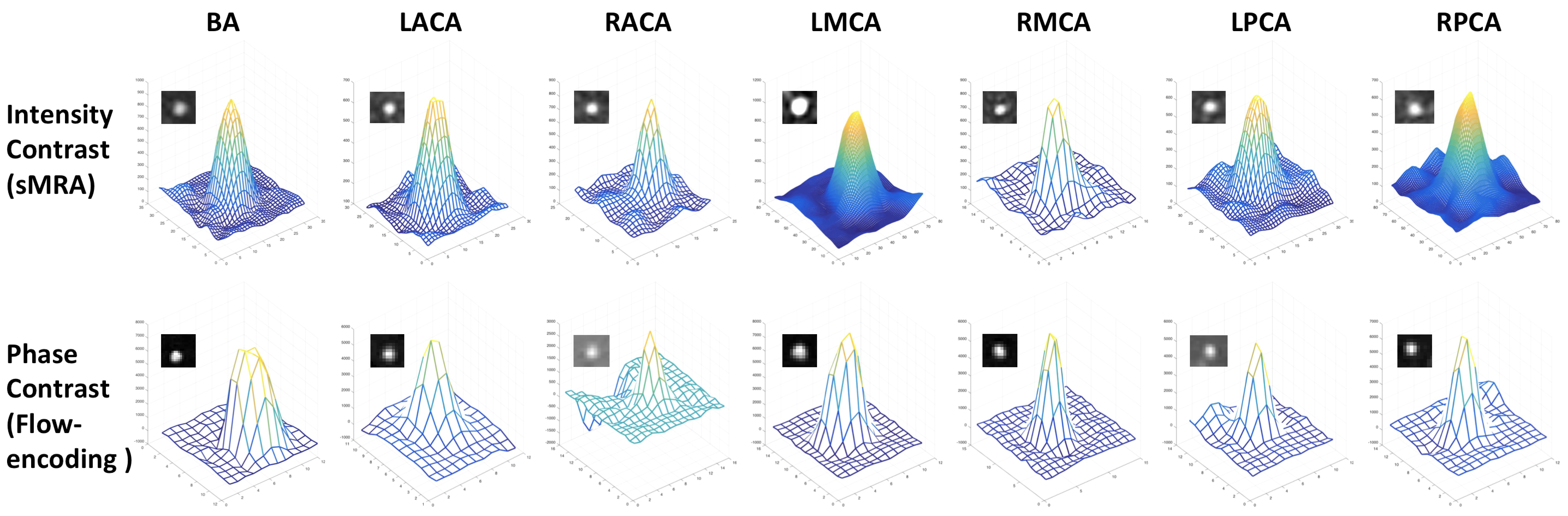
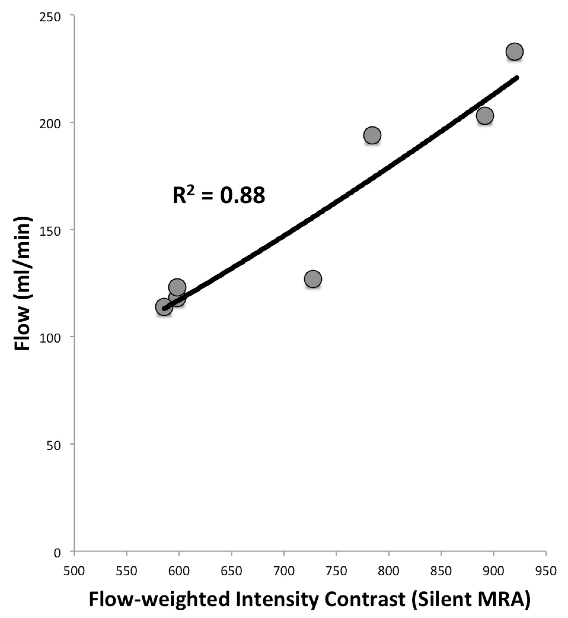
Compared to the TOF-MRA, Silent-MRA provided more details in arterial structure and more uniform intensities for tortuous vessels. In addition, the structural map from the Silent-MRA showed the gradient intensity that resulted from the flow-weighted information in the arterial regions (Figure 1). It is obvious that the Silent-MRA showed little signal loss compared to the TOF-MRA in the turns of the arteries. Extracting a region of interest (ROI) for each cerebral artery, the intensity distribution shown by the Silent-MRA closely agreed with the phase-contrast shape provided by the NOVA system (Figure 2, Table 1). Moreover, a linear correlation was found between the Silent-MRA signal intensity and the flow rates for all arteries (Figure 3). These results show that a Silent-MRA could provide a reliable flow-encoding arterial structure, and it would be useful in the diagnosis of arterial stenosis and its effect on blood flow.
CONCLUSION
Results show that the Silent-MRA images can provide a more accurate arterial structure than those from a traditional TOF-MRA, agreeing with previous studies1,2; moreover, the distribution of the flow-encoding Silent-MRA signal has a linear relationship with arterial flow rate. This preliminary study demonstrates the potential power of using the flow-encoding Silent-MRA signal for assessing complicated vascular disease.
Acknowledgements
No acknowledgement found.References
1. Irie R et al., Assessing Blood Flow in an Intracranial Stent: A Feasibility Study of MR Angiography Using a Silent Scan after Stent-Assisted Coil Embolization for Anterior Circulation Aneurysms, AJNR Am J Neuroradiol. 2015 May;36(5):967-70.
2. Shuang S et al., Follow-up assessment of coiled intracranial aneurysms using zTE MRA as compared with TOF MRA: a preliminary image quality study, Eur Radio., 2017, doi: 10.1007/s00330-017-4794-z.
3. Fujiwara Y and Muranaka Y, Improvement in visualization of carotid artery uniformity using silent magnetic resonance angiography, Radiol Phys Technol. 2017 Mar;10(1):113-120.
4. Takano N et al., Non-Contrast-Enhanced Silent Scan MR Angiography of Intracranial Anterior Circulation Aneurysms Treated with a Low-Profile Visualized Intraluminal Support Device, AJNR Am J Neuroradiol. 2017 Aug;38(8):1610-1616.
Figures