2916
Single breath-hold MR T1-mapping in the heart: comparison of hybrid MOLLI and MOLLI53Yu Chun-Yang1, Huang Teng-Yi2, and Chung Hsiao-Wen1
1National Taiwan University, Taipei, Taiwan, 2National Taiwan University of Science and Technology, Taipei, Taiwan
Synopsis
A hybrid MOLLI method that integrated saturation recovery with the classical inversion recovery sequence was proposed for quantitative T1 mapping in the myocardium within one single breath-hold. By replacing the second inversion pulse of the original MOLLI53 technique with a saturation pulse, the long recovery time could be alleviated in hybrid MOLLI, thereby allowing more images to be sampled from the T1 relaxation curve. Phantom and healthy subject experiments conducted in comparison with the classical MOLLI53 demonstrated that the proposed method was able to provide comparable image quality as well as precise T1 quantification in the myocardium.
Introduction
Alternations of myocardial T1 values have been shown to be related to disease states. Therefore, characterization of the native T1 values may provide useful information to detect and assess cardiomyopathies. In terms of T1 measurements, inversion recovery methods such as MOLLI are widely used clinically but are subject to magnetization transfer effects (1), whereas saturation recovery methods including SASHA are potentially more accurate but are prone to noise influences (2). The purpose of this study is therefore to propose a new hybrid method combining inversion and saturation recovery and to investigate the precision of T1 measurements on both phantoms and healthy subjects.Materials and Methods
The hybrid MOLLI method proposed in this study is composed of an inversion pulse after which five images are acquired, followed by a saturation pulse after which six images are acquired (Fig.1). Compared with the original MOLLI5(3s)3 design (two inversion pulses separated by 3 seconds, with five and three images acquired after each inversion pulse, respectively), there is no need to wait for full recovery of the magnetization, hence allowing more images to be sampled for the T1 relaxation curve (3). A phantom consisting of eight bottles of MnCl2 solution was imaged at 3T using the original MOLLI5(3s)3 and the hybrid MOLLI, both repeated twice to assess reproducibility. The T1 values estimated were compared against inversion recovery turbo spin-echo as the gold standard. Simulated ECG was varied from 50 bpm to 90 bpm to assess the effect of heart rate variability. In addition, twelve healthy subjects were imaged using both MOLLI5(3s)3 and hybrid MOLLI in a breath-hold. The measurements were again repeated twice to assess reproducibility. T1 was estimated on a pixel-by-pixel basis using nonlinear least square error fitting in MATLAB. Myocardial T1 values measured from selected regions-of-interest were compared.Results
Nonlinear fitting of the data to the signal model was successful (Fig.1). Both MOLLI53 and hybrid MOLLI showed T1 estimation with good precision, although underestimation was found when compared with inversion recovery turbo spin-echo measurements in the phantom (Fig.2). Figure 3 shows one representative slice of T1 maps acquired using MOLLI53 and hybrid MOLLI, respectively. It is seen that both techniques were able to accomplish T1 mapping in a single breath-hold, and yielded quantitative values very similar to each other. Regions-of-interest measurements from the myocardium also showed comparable precision at the level of about 3% errors for MOLLI53 and hybrid MOLLI (Fig.4).Discussion
The results from our healthy subject study suggest that the hybrid MOLLI technique combining saturation recovery and inversion recovery is an effective alternative to the widely used MOLLI5(3s)3 sequence for quantitative T1 mapping in the myocardium. The use of saturation pulse instead of a second inversion pulse alleviated the need of long recovery periods as in the conventional MOLLI53 sequence (3), allowing more images to be acquired than conventional MOLLI5(3s)3. In the case of patients with arrhythmia, it is expected that the hybrid MOLLI method may provide advantages by sufficiently sampling the T1 relaxation curve.Conclusion
The hybrid MOLLI technique proposed in this study has been shown to yield precise T1 mapping in a single breath-hold, with quality comparable to that shown in the conventional MOLLI53 method on phantoms and healthy subjects.Acknowledgements
No acknowledgement found.References
- Kellman P, Hansen MS. T1-mapping in the heart: accuracy and precision. J Cardiovasc Magn Reson 2014;16:2.
- Kim PK, Hong YJ, Im DJ, et al. Myocardial T1 and T2 Mapping: Techniques and Clinical Applications. Korean J Radiol 2017;18:113-131.
- Roujol S, Weingärtner S, Foppa M, et al. Accuracy, Precision, and Reproducibility of Four T1 Mapping Sequences: A Head-to-Head Comparison of MOLLI, ShMOLLI, SASHA, and SAPPHIRE. Radiology 2014;272:683-689.
Figures
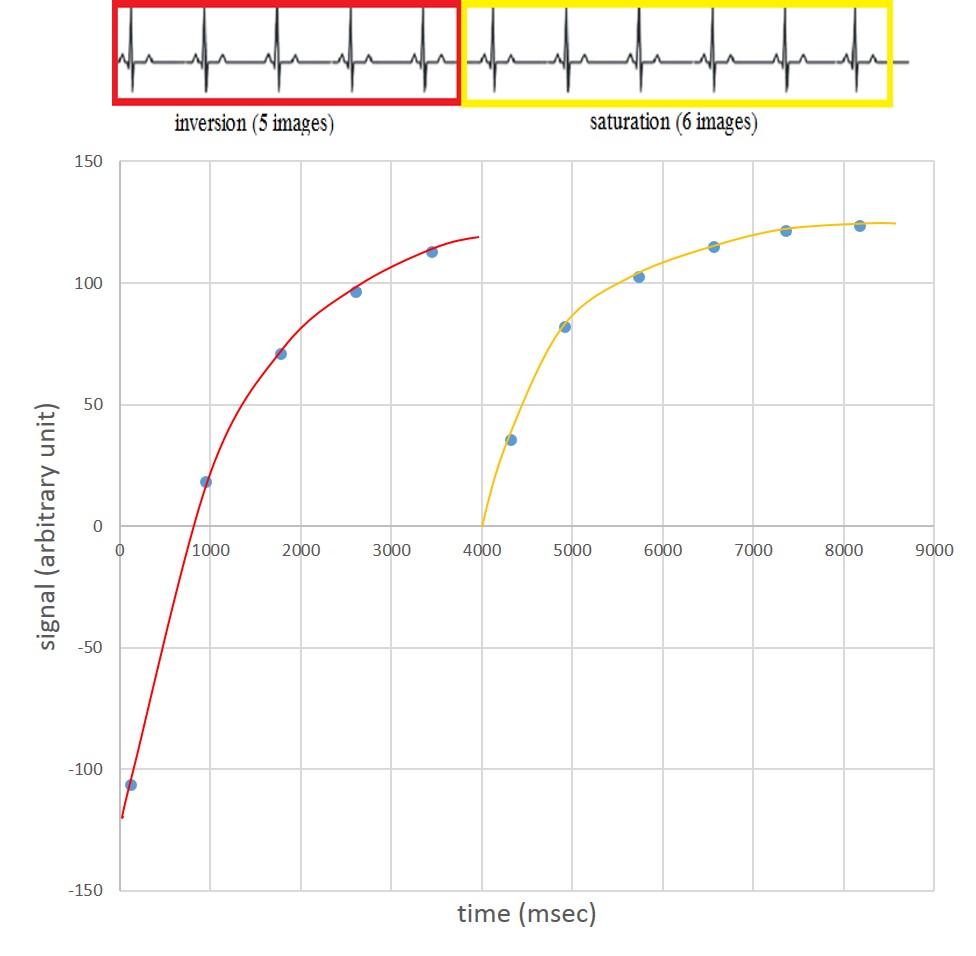
Figure 1. The schematic of hybrid MOLLI proposed in this study. Five images
corresponding the same cardiac phase are acquired after the first inversion
pulse, followed immediately by a saturation pulse after which six images are
acquired. Experimental data demonstrating the goodness of fit are also shown.
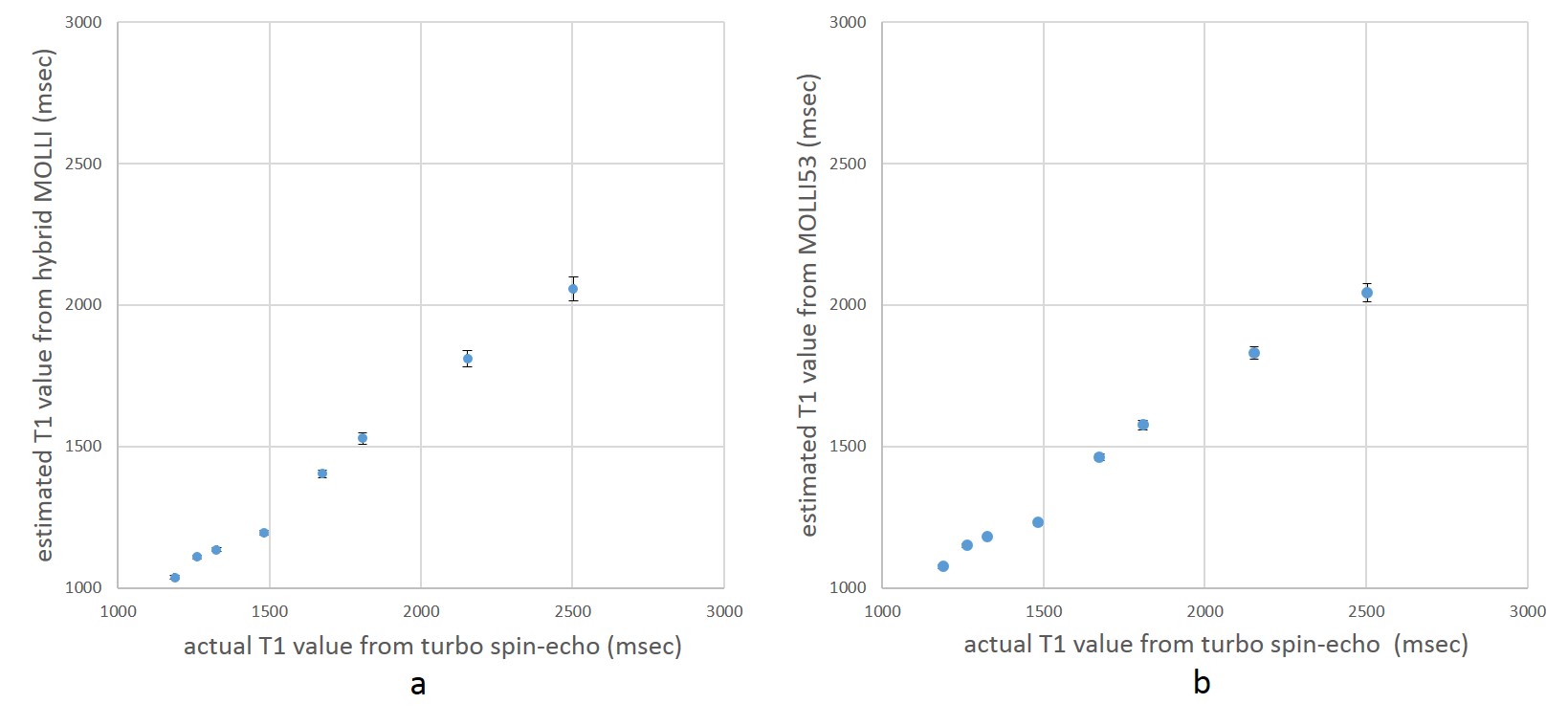
Figure 2. T1 values estimated using (a) hybrid MOLLI and (b) MOLLI53 from the MnCl2
phantom plotted against those measured using inversion recovery turbo spin-echo.
Both MOLLI methods showed T1 underestimation but with similar good precision.
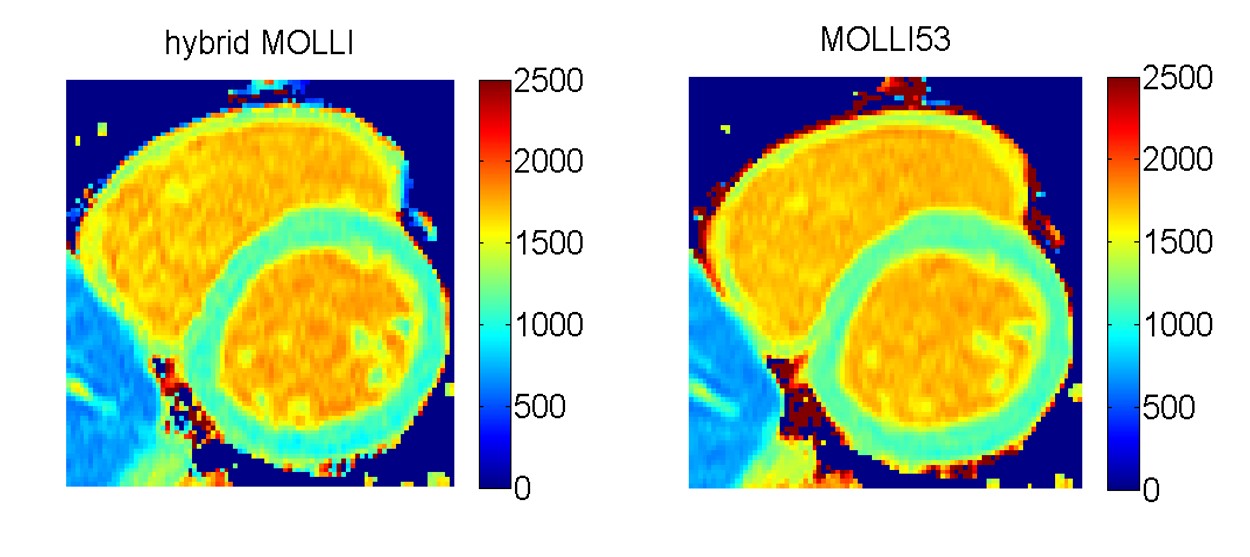
Figure 3. Representative slice of T1 maps acquired in one single breath-hold using
MOLLI53 (right) and hybrid MOLLI (left), which are visually comparable to each
other.
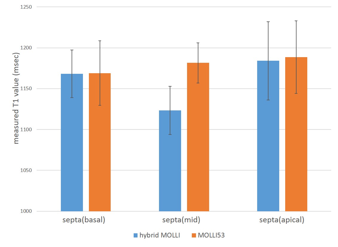
Figure 4. T1 values estimated from three regions-of-interest within the myocardium
using hybrid MOLLI (blue) and MOLLI53 (orange), showing numerical T1 values in
good agreement with literature values and comparable precisions at the level of
about 3% errors for the two techniques.