2912
Fast and precise myocardial T1 mapping using a segmented golden angle radial MOLLI sequence with bSSFP readout1Radiology, UCLA, Los Angeles, CA, United States, 2UCLA, Los Angeles, CA, United States
Synopsis
Among the various myocardial T1 mapping sequences developed, the radial variants of the MOLLI acquisitions (raMOLLIs) are promising. As raMOLLIs can decrease the acquisition time down to a few heartbeats while keeping good T1 estimation precision due to a large number of images reconstructed along the T1 relaxation recovery curve. The previous raMOLLIs use FLASH readout due to the sensitivity of bSSFP readout to image artifacts. As bSSFP readout has high SNR, a variant of radial MOLLI with bSSFP readout was developed to ensure accurate and precise myocardial T1 mapping by using segmented golden angle radial acquisition.
Background
Myocardial T1 mapping is an emerging technique used to assess diffuse myocardial fibrosis. Among the various myocardial T1 mapping sequences developed, the radial variants of the MOLLI acquisitions (raMOLLI) (1,2) are promising. As raMOLLIs can decrease the acquisition time down to a few heart beats, while keeping good T1 estimation precision due to a large number of images reconstructed along the T1 relaxation recovery curve. T1 estimations can also be insensitive to heart rate variations (1). However, the current developed raMOLLI sequences are all based on the FLASH readout, and not using the benefit of the bSSFP readout, which has a much higher signal to noise ratio (SNR) compared to the FLASH readout. The main reason may be that, in gold angle radial acquisition, bSSFP readout is highly subject to eddy current artifacts. In this work, we sought to develop a variant of raMOLLI sequence with bSSFP readout, which can eliminate the image artifacts to generate accurate and precise myocardial T1 maps.Methods
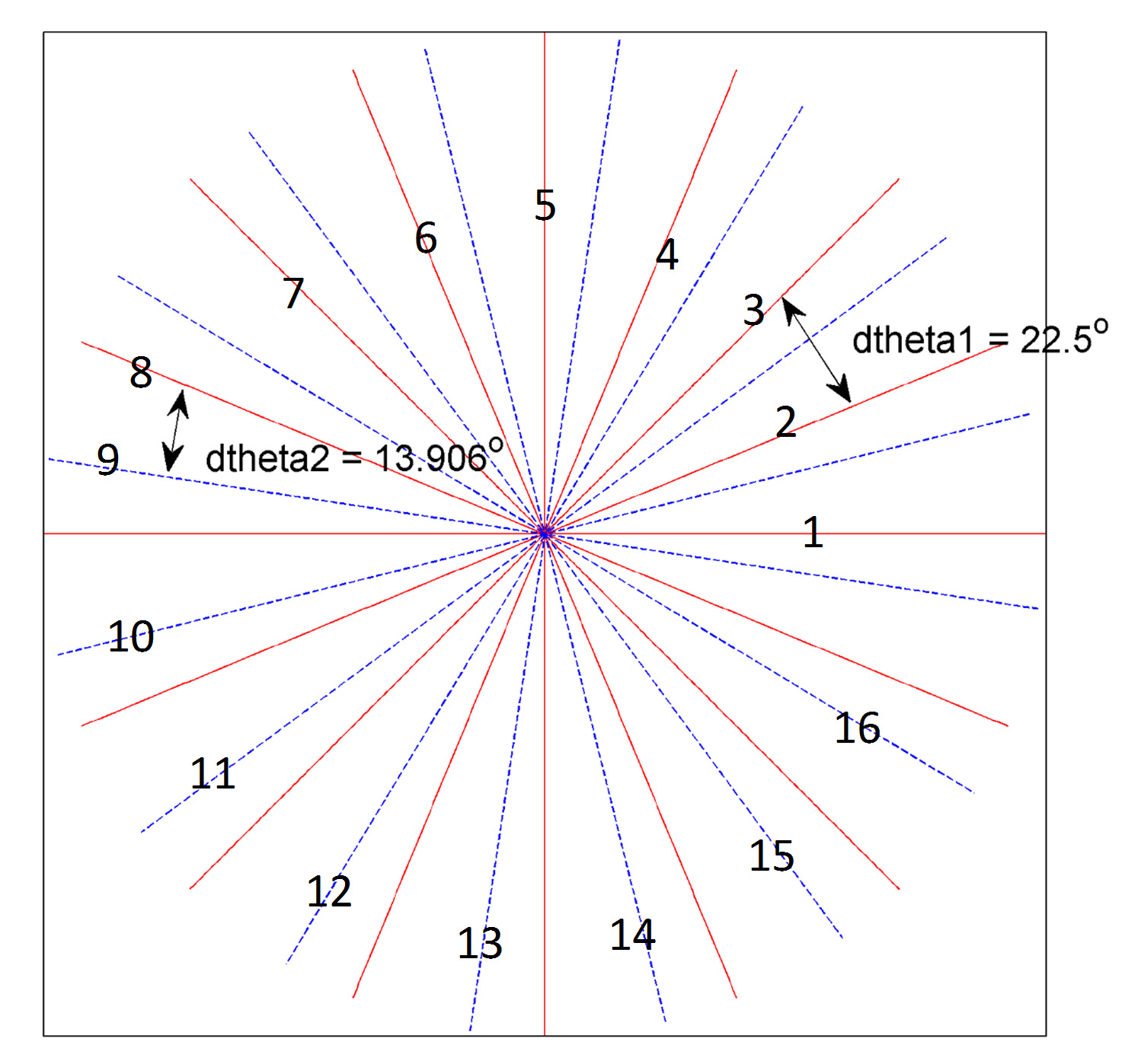
The raMOLLI with bSSFP readout was implemented on a 1.5 MRI scanner (Avanto Fit Siemens Healthcare; Erlangen, Germany). Similar to a previous study (1), after inversion, 5 shots of 80 radial spokes were acquired. Two different acquisition schemes were tested: golden angle (GA, 111.246° angle increase) and our proposed segmented golden angle (sGA) radial acquisition. The detail of segmented golden angle radial acquisition is illustrated in Figure 1, where every 8 spokes form a group and were evenly distributed in all 8 segments. During k-space acquisition, the radial angle increase was either 22.5º or 13.906 º (= 22.5ºx Golden ratio) depending on whether the next radial line belongs to the same group or next group. By this approach, the eddy current image artifacts can be reduced, as the radial angle increase was no more than 22.5º. For each shot, 10 images were reconstructed using a view sharing method and a KWIC filter, which contained 5 layers, with 8*(1,2,3,5,7) =16, 24, 40 and 56 radial spokes in each layer for our sGA approach, and 8, 13, 21, 34, 55 radial spokes in each layer for GA approach. Under-sampled data were then reconstructed using a compressed sensing algorithm with total variation regularization. Bloch equation simulation with slice profile correction (BLESSPC) T1 estimation (3) was used to generate T1 maps based on the reconstructed images.
Eight gel phantoms were used. GA based radial MOLLI was acquired with a flip angle (FA) = 35º, while sGA based radial MOLLI was acquired with FA = 25º, 35º and 70º at a simulated heart rate of 60 bpm. Reference T1 for each gel phantom was determined by a standard inversion-recovery spin echo technique. The proposed radial MOLLI with bSSFP readout was also evaluated in one healthy volunteer to demonstrate its ability to remove image artifacts compared GA based radial MOLLI.
Results
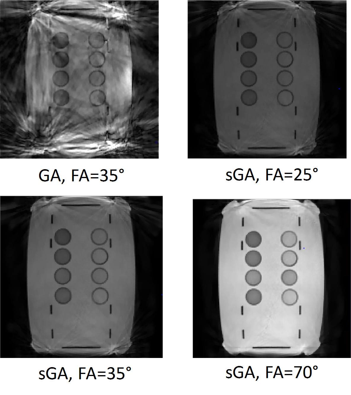
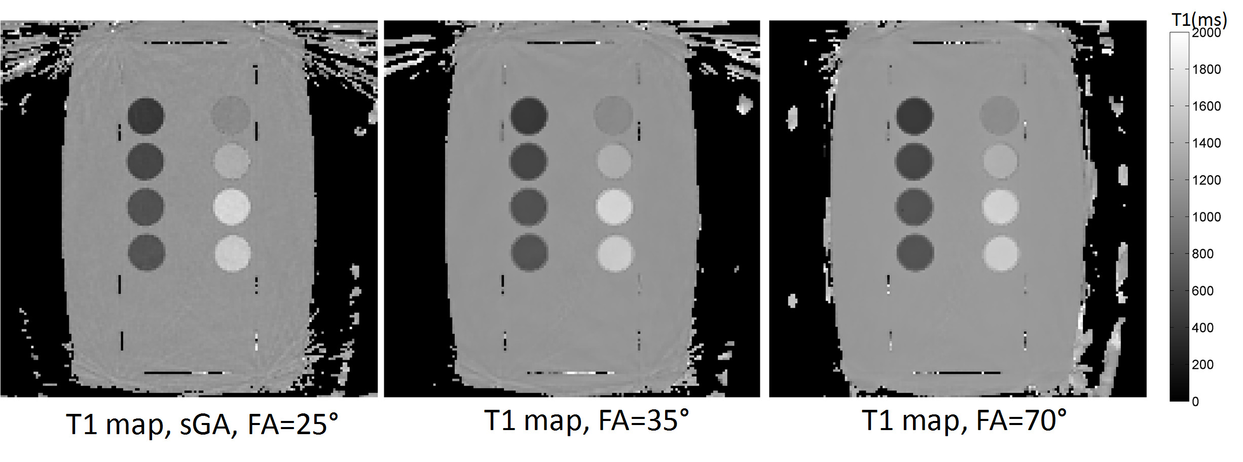
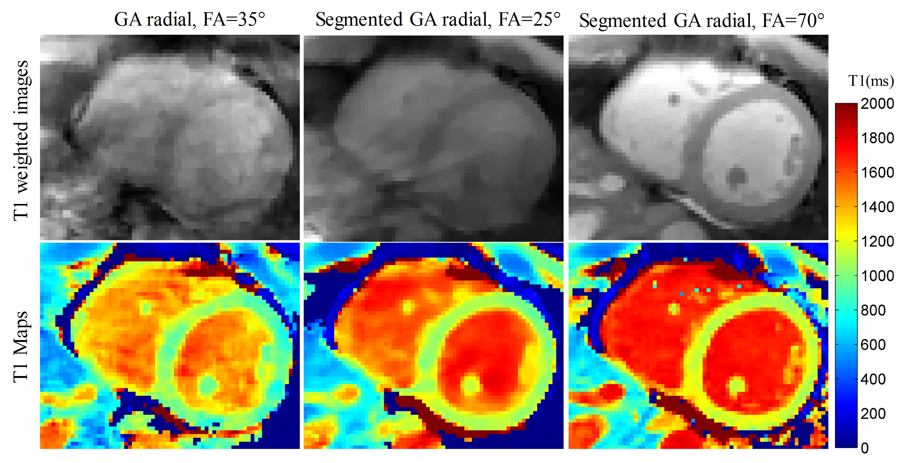
Figure 2 shows one of the reconstructed T1 weighted image using GA radial-MOLLI and sGA radial-MOLLI with bSSFP readout and at different flip angles in phantom studies. It shows that our proposed sGA based radial-MOLLI can reduce the image artifacts greatly, and with higher flip angle (FA=70º), the image artifacts can further be eliminated, resulting in T1 maps with less artifacts and noise, as shown in Figure 3. T1 estimation result correlate will with the reference T1 values using proposed radial-MOLLI and FA=70º (R2 = 0.998) and the average absolute T1 estimation error is 3.4%±1.9%. Figure 4 shows one of the reconstructed T1 weighted image using GA radial-MOLLI and sGA radial-MOLLI with bSSFP readout and at different flip angles acquired in a healthy volunteer. Similar to phantom results, segmented gold angle based radial-MOLLI with FA=70º generate the best quality T1 weighted images and T1 map.Discussion and conclusion
This work demonstrates the benefit of using segmented gold angle radial-MOLLI with higher flip angle bSSFP readout to reduce image artifacts for accurate and precise myocardial T1 mapping at 1.5T. The idea of using small angle increase by using tiny gold angle has been proposed to reduce similar artifacts for radial acquisition with bSSFP readout. However, tiny gold angle requires a minimal number of radial spokes to ensure relatively uniform under-samples. In radial-MOLLI, the first layer of KWIC filter uses only 8 spokes, which doesn’t satisfy the tiny gold angle condition, and therefore tiny gold angle cannot be applied in radial-MOLLI. Further studies are warranted to study higher flip angle can help to reduce image artifacts.Acknowledgements
NoneReferences
1. Marty B, Coppa B, Carlier PG. Fast, precise, and accurate myocardial T 1 mapping using a radial MOLLI sequence with FLASH readout. Magn. Reson. Med. 2017. doi: 10.1002/mrm.26795. 2. Gensler D, Mörchel P, Fidler F, Ritter O, Quick HH, Ladd ME, Bauer WR, Ertl G, Jakob PM, Nordbeck P. Myocardial T1: Quantification by Using an ECG-triggered Radial Single-Shot Inversion-Recovery MR Imaging Sequence. Radiology 2015;274:879–887. 3. Shao J, Liu D, Sung K, Nguyen K-L, Hu P. Accuracy, precision, and reproducibility of myocardial T1 mapping: A comparison of four T1 estimation algorithms for modified look-locker inversion recovery (MOLLI). Magn. Reson. Med. 2017;78:1746–1756.Figures