2883
Dynamic field map correction based on reversed-gradient design for non-Cartesian single-shot fast fMRI1Dept. of Radiology, Medical Center - University of Freiburg, Freiburg, Germany
Synopsis
A dynamic field map correction technique based on reversed-gradient design is introduced to non-Cartesian single-shot fast fMRI to correct the off-resonance artifacts. The field map estimated from dual-TE GRE scan could not capture that from field drift and eddy currents, so the off-resonance artifacts could not be corrected completely. This technique acquires two images with reversed slow-encoding directions in each time frame, which is generally used in EPI, and updates field map iteratively based on conjugate gradient reconstruction. After correction, the off-resonance artifacts are significantly reduced.
Introduction
Fast functional MRI (fMRI) plays an important role in investigating the working mechanism of human brain. One challenge for fast fMRI using single-shot trajectories is that it is prone to off-resonance artifacts due to the relatively long readout duration. Off-resonance artifacts can be corrected to some extent by modeling the additional signal dephasing calculated from a separately acquired static field map1, estimated for example from two or more gradient echo (GRE) reference scans with different echo time (TE). However, this static field map may be inaccurate with respect to a practical fMRI scan because many factors, such as field drift, eddy currents, and motion, could introduce additional time-dependent magnetic fields, leading to an incomplete correction for off-resonance artifacts2. Several dynamic field map correction methods have been proposed, such as reversed phase-encoding method2, jittered echo time method3, but all of them are designed for echo-planar imaging (EPI) sequence. This study extends the reversed phase-encoding method to model-based image reconstruction for non-Cartesian single-shot fast fMRI to correct field map dynamically.Methods
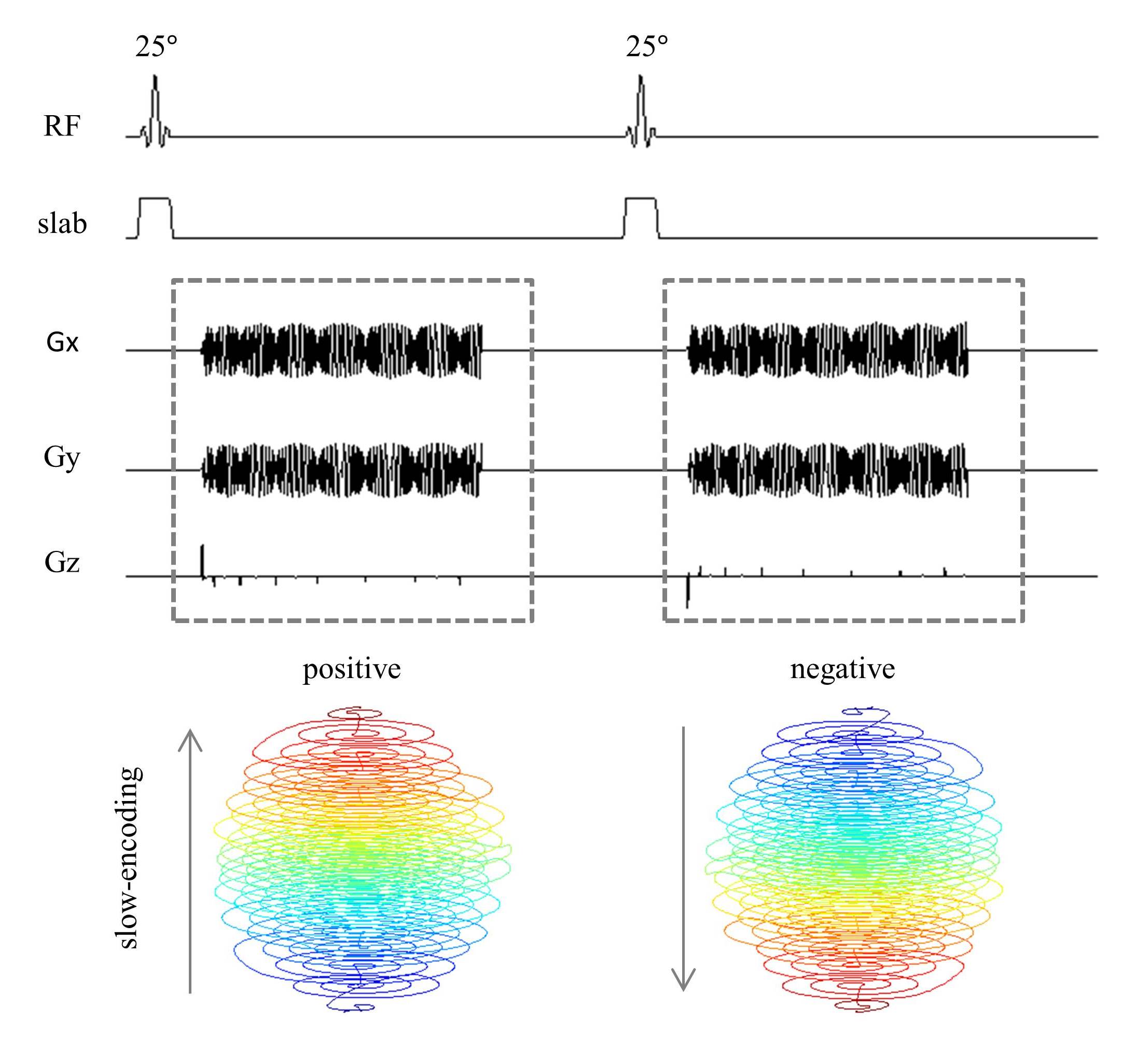
A phantom was measured on a 3T MR scanner (Magnetom Prisma, Siemens, Erlangen, Germany) with a 64-channel head coil. Dual-echo GRE reference images (TR 500ms, TE1 4.92ms, TE2 7.38ms, flip angle 70deg, spatial resolution/FOV 3×3×3/192×192×192mm) were acquired to calculate coil sensitivity maps and original field maps. A structure image with 1×1×1mm resolution was collected as reference. The 3D single-shot fMRI sequence, MREG (TR 100ms, flip angle 25deg, spatial resolution/FOV 3×3×3/192×192×192mm) with stack-of-spirals trajectory, was used4. This was alternated with an identical trajectory, but with reversed gradient polarities, in successive excitations, as shown in Fig.1.
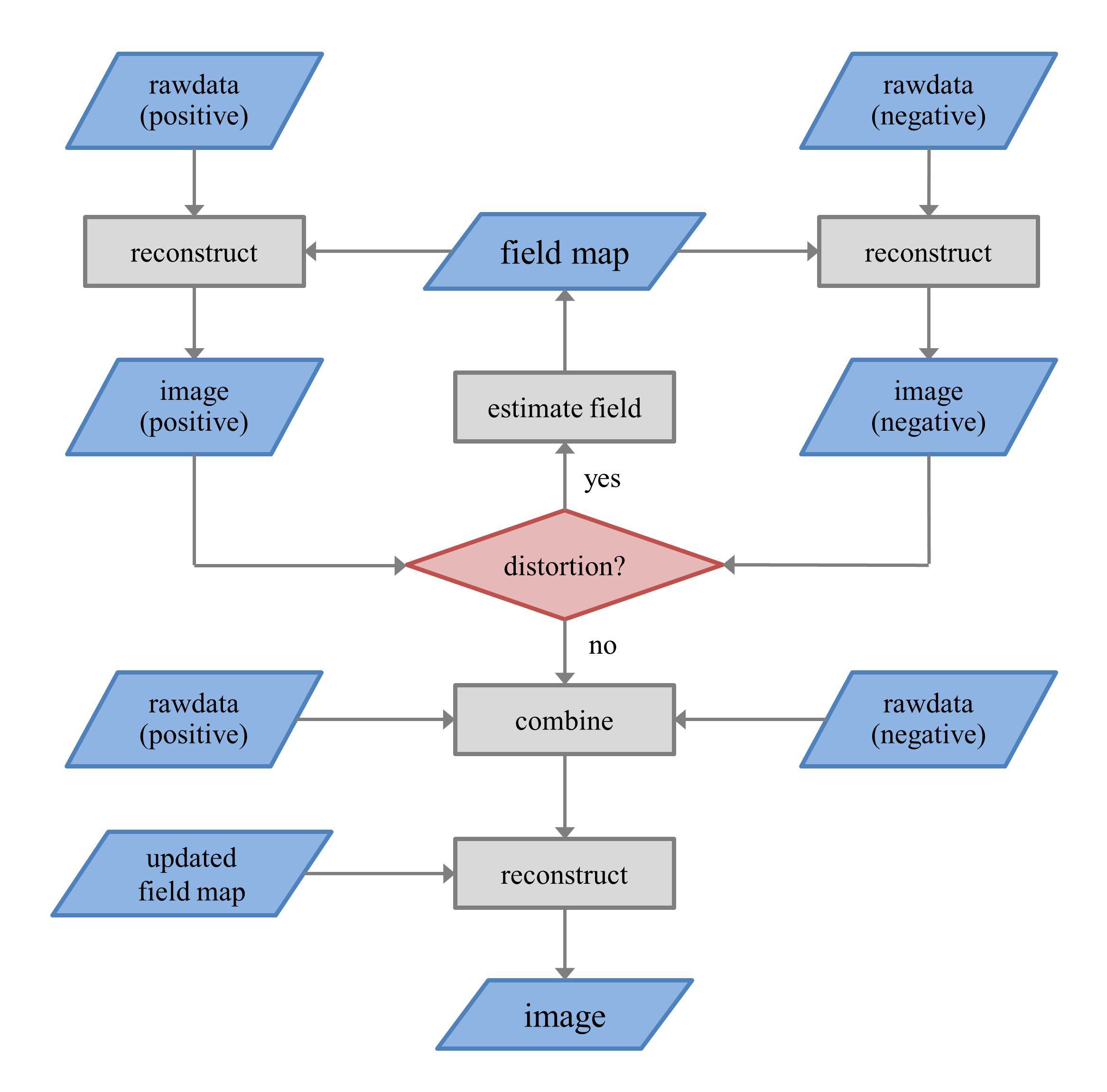
The reconstruction is based on nonlinear conjugate gradient SENSE (CG-SENSE) algorithm. Here, two excitations are regarded as one time frame. The pipeline of dynamic field map correction is as following (Fig.2): (1) reconstruct the images from two shots separately using the current field map estimate (using the statically acquired field map as the initial estimate); (2) estimate distortions between the two reversed-gradient images by non-linear coregistration and calculate the field map that best explains these distortions; (3) update the field map by adding the estimated field map to the current field map; (4) repeat step 1-3 until there is no distortion mismatch between the two reconstructed images; (5) reconstruct images using the combined datasets and updated field map. This procedure is implemented at each time frame to correct off-resonance artifacts dynamically.
Results
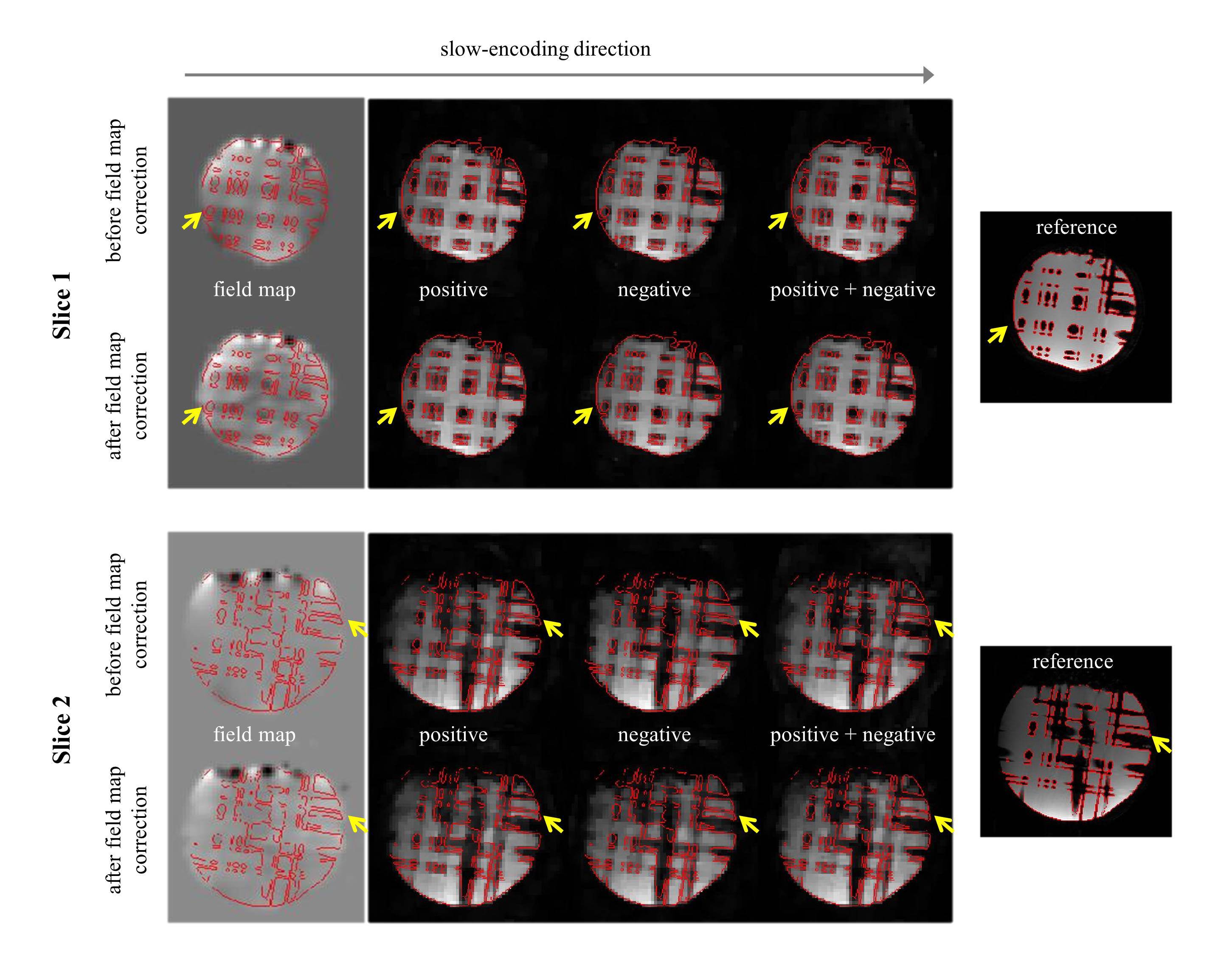
Fig.3 presents the field maps and reconstructed images before and after field map correction in the first time frame. With the original field map, the distortion artifacts relative to reference (the red edge is extracted from reference) still exist in the positive and negative images along the slow-encoding direction. It is reduced by joint reconstruction (positive + negative), but there are still residual artifacts in the mismatch areas. The correction results in an altered field map estimate, especially in the mismatch areas. This correction successfully removes the mismatch between positive and negative images. With joint reconstruction, the distortion is further corrected comparing to the reference.Discussion
The results show that the field map obtained from the dual-TE GRE sequence could not completely correct image distortions in subsequent fMRI acquisitions. The field map correction technique removes off-resonance artifacts in two aspects: (1) correcting the field map by reversed-gradient design which minimizes the distortion mismatch between two images; (2) removing the distortion by joint reconstruction which, in essence, is a compromise of the opposite distortion artifacts. As an accurate field map is important in correcting distortions, the joint reconstruction could further remove the distortion even when the field map is not so accurate.Conclusion
This study introduces reversed-gradient design to non-Cartesian single-shot fast fMRI and CG-SENSE reconstruction and shows that it is feasible to correct field map dynamically to take into account factors such as field drift, eddy currents…Acknowledgements
This work was supported by the DFG Koselleck grant He 1875/28-1, the cluster of excellence EXC-1086 BrainLinks-BrainTools from the DFG, and the China Scholarship Council (CSC).References
1. Sutton, Bradley P., Douglas C. Noll, and Jeffrey A. Fessler. Fast, iterative image reconstruction for MRI in the presence of field inhomogeneities. IEEE transactions on medical imaging. 2003; 22(2): 178-188.
2. Jezzard P, Barnett A S, Pierpaoli C. Characterization of and correction for eddy current artifacts in echo planar diffusion imaging. Magnetic resonance in medicine. 1998; 39(5): 801-812.
3. Andersson, Jesper LR, Stefan Skare, and John Ashburner. How to correct susceptibility distortions in spin-echo echo-planar images: application to diffusion tensor imaging. Neuroimage. 2003; 20(2): 870-888.
4. Assländer, Jakob, et al. Single shot whole brain imaging using spherical stack of spirals trajectories. Neuroimage. 2013; 73: 59-70.
Figures