2879
Improved MR Neurography of Brachial Plexus at 3.0 T with iMSDE and DIXON methods1Philips Healthcare, Beijing, China, 2Beijing Jishuitan Hospital, Beijing, China
Synopsis
In this study we developed a sequence for neurography with robust fat and blood suppression for increased conspicuity of nerves. Improved Motion-Sensitized Driven-Equilibrium Preparation (iMSDE) was applied to null blood and lymphatic motions. DIXON TSE readout with long TE was used to generate water images representing nerves, in brachial plexus where other fat suppression pulses were challenged by B0 and B1 complexities. Preliminary test in brachial plexus showed its advantages and stability.
Purpose
Chronic Inflammatory Demyelinating Polyneuropathy presents a chronic progressive or a relapsing-remitting disorder, comes with prolonged the T2 relaxation time in the nerve involving fascicular enlargement(1). Several MR Neurography methods have been developed to selective depict of peripheral nerves, such as in brachial plexus and lumbar plexus. Recently developed (2,3) improved Motion-Sensitized Driven-Equilibrium Preparation (iMSDE) pre-pulse was applied for venous signal suppression, and showed robust plexus visualization (3D Nerveview, Philips). Optimized low-refocusing-flip-angle turbo spin echo sequence combined is used to generate the high signal image without geometry distortions. A fat saturation pulse (for example, STIR or SPIR) is required, due to long T2 effect of fat. However, fat saturation pulses’ performance rely on the B0 homogeneity which is rather challenging in regions such as brachial plexus. It has been investigated to use multiecho TSE modified Dixon (mDixon) to obtain robust fat suppression (4). And mDixon techniques also intrinsically suppresses blood signal, with the TSE acquisition.
While 3D Nerveview has difficulty in fat suppression and mDIXON TSE has its sub-optimal in blood suppression, we developed 3D mDIXON TSE with iMSDE pre-pulse. This strategy applies two point Dixon acquisition and a strong blood suppression to achieve higher nerve signal contrast.
Methods
This study was approved by institutional research board; healthy volunteers were recruited for image comparison. MR neurography images were collected on a 3.0T MR Scanner (Ingenia, Philips Healthcare, Best, the Netherlands). 3D refocusing-flip-angle-controlled TSE with iMSDE and fat saturation preparation pulses(3D Nerveview) images, and the novel 3D TSE DIXON MSDE sequence were performed.
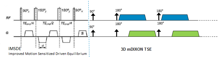
3D multi-shot turbo spin-echo (TSE) sequence is modified to acquire both near-in-phase and near-out-of-phase echoes, as required for chemical shift (DIXON) reconstruction. The iMSDE preparation pulse, set to generate optimal black-blood effect for nerve imaging, is added in front of DIXON TSE acquisition to avoid flow liquid signal from blood.
Image resolution is 1.2×1.2×2mm^3, with corresponding acquisition matrix 292×334, TR 2s, TE set 180ms and TSE factor 51, and compressed SENSE with acceleration factor 7 is also applied to reduce scan time to 5min 26s, equivalent to 3D Nerveview with STIR fat suppression at 5min 32s.
Figure 1 demonstrated the 3D TSE DIXON MSDE sequence, with iMSDE blood suppression ahead of two series of TSE image acquisition at shifted echo times.
Results
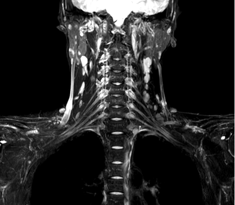
Figure 2 shows the clinically applied 3D Nerveview image.
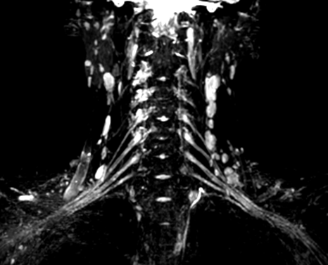
Figure 3 shows images from the 3D DIXON TSE MSDE sequence.
The 3D DIXON TSE MSDE sequence successfully combined the fat suppression and venous suppression features; and nerve visualization were significantly improved. Significant better fat suppression was obtain in 3D DIXON TSE MSDE than in 3D Nerveview.
Conclusion and Discussion
The multiecho 3D TSE-mDixon sequence provides robust fat and blood suppression, resulting in increased conspicuity of the nerves, and scan time is affordable in clinical settings to achieve superior MR neurography at the brachial plexus. However, scan time is longer for the mDIXON TSE multi acquisition method, the multi-echo mDIXON TSE in (4) could potentially shorten scan time in addition to compressed SENSE acceleration.
However, there are still some residual blood signals in 3D DIXON TSE MSDE images, which might relates to the directional suppression of iMSDE feature. The blood flowing in diagonal direction kept some signal in the FOV. However, the iMSDE eliminated majority part of blood signal and destroyed the formation of vessel, made it easier to distinguish between vascular anatomy with nerves.
Further research will be carried out to validate this sequence with positive findings among patients.
Acknowledgements
No acknowledgement found.References
- Latov N, Nat Rev Neurol. 2014 Aug;10(8):435-46.
- Wang J, et al. MRM 2007; 58(5):973-81
- Dong L, et al. ISMRM. 18 2010:1274
- Wang X, et al. MR Neurography of Brachial Plexus at 3.0 T with Robust Fat and Blood Suppression, Radiology, 2017
Figures