2875
Quantification and management of MR image spatial accuracy for applications in radiation therapyTeo Stanescu1, Joanne Moseley2, Callum Moseley2, Mostafa Shahabi2, and David Jaffray1
1Medical Physics, Radiation Medicine Program, Princess Margaret Cancer Centre, University Health Network & University of Toronto, Toronto, ON, Canada, 2Medical Physics, Radiation Medicine Program, Princess Margaret Cancer Centre, University Health Network, Toronto, ON, Canada
Synopsis
An automated imaging pipeline was developed and validated to handle the management of MR image spatial accuracy with a focus on applications for radiation therapy (RT). Protocol enforcement was implemented to accept/reject datasets based on expected clinical sequence parameters. System and patient related image spatial distortions were quantified using numerical simulations and measurements. Vector field maps were rendered and stored for automatic filtering and correction of patient MR images. Data and process monitoring was enabled via a web application. The imaging pipeline was deployed clinically to automatically validate patient image data required for RT planning and in-room MR-guided treatment delivery.
Introduction
Radiation therapy requires a high degree of MR image spatial accuracy since images are directly used to contour, plan and guide the treatments of cancerous targets. Knowing the exact topography of the target is critical for ensuring safe delivery, sparing neighboring healthy organs at risk, and maximizing curative intent. Methods are required to quantify all issues affecting the spatial accuracy of images. Further, to make the methods practically feasible and efficient in a clinical environment, analysis and process automation are highly desirable to handle strenuous demands regarding image data - i.e., volume, near real-time availability, paramount safety and excellent overall quality.Methods
A harmonic analysis (HA) based on solving an inner and outer Dirichlet boundary value problem (BVP) using finite element methods (FEM) was developed and validated to quantify 3D system-related distortion fields, which are due to B0 inhomogeneities and gradient nonlinearities. The BVP consists of solving the Laplace equation with boundary conditions (BCs) for the inside and outside of an arbitrarily-shaped domain of interest. The BC was defined as a continuous shape function representing the distortion vector field on the domain’s surface. Solving the BVP, the 3D vector fields were generated inside the entire domain and beyond the surface, up to a given distance and error tolerance. The HA fields were compared with measured fields obtained with a full field of view (FOV) grid-type linearity phantom. For practicality reasons, an HA-driven hollow cylindrical phantom was also designed and built to map full MR FOVs for this study. To note, the size of the phantom was significantly smaller than the MR FOV – the outer BVP-derived field provided the distortions at the periphery of the MR FOVs. To quantify the patient-induced distortions, a GPU-based magnetic field analysis relying on finite difference methods (FDM) was developed and implemented for imaging protocol optimization. The analysis requires CT/MR input image data and tissue susceptibility values to compute local distortions (ppm/mm) by taking into consideration the scanner field and readout gradient values. The automation and imaging pipeline were developed in a Python environment with a software architecture comprising of two virtual machines (VMs) – i.e., image processing, numerical analysis, storage of analytics in a relational database, hosting of web application with user login and dashboards for data display, monitoring, reporting and audit (see Figure 1).Results and Discussion
The harmonic analysis was validated for multiple quadratic volumes and full MR imaging FOVs – the residual errors when compared to the reference fields were lower than the measurement errors (1 mm) – see Figure 2. The cylindrical phantom and HA were used to collect data for the correction of patient data. The HA-derived fields were stored and uniquely tagged for each imaging protocol for patient data correction. The FDM modeling for the patient-induced distortions was found in excellent agreement with phantom data and simulations were performed to establish specific error levels for all RT treatment sites (location, magnitude). The automated imaging pipeline was implemented on the hospital network to receive DICOM images and sort them based on content and relevance to the pipeline analysis. Once sorted, analysis processes were automatically triggered and results/analytics sent via a dedicated API to an SQL database. A web server and application were configured to parse the database, extract analytics and display them when available via customized dashboards. Secure login was also implemented for multi-user access and multiple MR scanners (institutions and sites). Alternative configurations with both VMs hosted on Amazon Web Services (AWS) or hybrid, hospital network and AWS, were also tested.Conclusion
MR image distortions relevant to radiation therapy were quantified using numerical methods based on harmonic analysis and magnetic field computations. The research work was implemented clinically using a software architecture designed to allow for the automatic management of image data (sanity check, correction, reporting). The work was performed under the MR-guided adaptive radiotherapy project at our institution, which has at its core a hybrid MR/linear accelerator system designed for the acquisition of MR images in the treatment room and their use for treatment planning and radiation guidance.Acknowledgements
The work was supported by funding from the Princess Margaret Cancer Foundation.References
No reference found.Figures
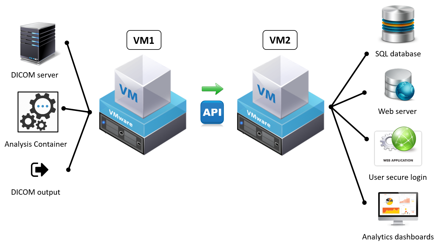
Automated
imaging pipeline for the management of MR image distortions. Two virtual
machines host all processes required for the automated analysis and data-flow. The
analysis container is modular and can run single or multiple (serial/parallel)
processes on same dataset. Corrected image sets are forwarded to DICOM-destinations
for RT planning and in-room guidance. Process analytics/logs are transferred
through a dedicated API into a relational database, and subsequently mined for
further analysis and visualization via a web application. Process feedback is
provided via email to MR users near real-time (~1 min) to inform about the quality
and status of acquired image data.
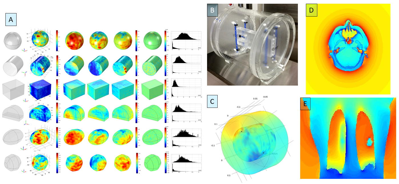
A)
Summary results of the harmonic analysis for multiple geometries (sphere,
cylinder, cuboid, D-shape, ellipsoid, and Reuleaux 9-gon); B) Phantom built to
provide proof-of-concept for HA and to collect 3D vector field data for the
automated patient image data correction on the imaging pipeline; C)
Example 3D FEM results for the combined Dirichlet problem corresponding to the
inner and outer regions of the cylindrical phantom; D) and E) Sample
results of the FDM-based magnetic field computations - susceptibility-induced
effects (ppm) are shown for a brain mets and a lung patient, respectively.