2866
Correcting for contrast differences across 3D T1 acquisitionsSean N Hatton1,2, Donald J Hagler1,3,4, Joshua Kuperman2,3,4, William S Kremen1,2, and Anders M Dale1,2,4,5
1Department of Psychiatry, University of California, San Diego, La Jolla, CA, United States, 2Center for Behavior Genetics of Aging, University of California, San Diego, La Jolla, CA, United States, 3Department of Radiology, University of California, San Diego, La Jolla, CA, United States, 4Center for Multimodal Imaging and Genetics, University of California, San Diego, La Jolla, CA, United States, 5Department of Neurosciences, University of California, San Diego, La Jolla, CA, United States
Synopsis
The aim of this study was to correct volumetric differences between images acquired with different MRI parameters. We scanned six subjects on the same 3.0T MRI scanner using different T1-weighted imaging sequences. Images were corrected for gradient warping and intensity inhomogeneity, then we applied a novel white matter intensity scaling and a voxel-wise image intensity normalization process. The correction improved the goodness of fit, precision and accuracy of the volumetric segmentation of the target image to each test sequence (typically < 1% difference). This procedure is particularly effective for voxel-wise segmentation techniques over surface-based approaches.
INTRODUCTION
Estimations of brain structure measurements may vary due to changes in magnetic resonance imaging (MRI) scanner parameters that affect tissue contrast profiles. The aim of this study was to correct for such sequence-specific contrast properties by using a two-stage image normalization process.METHODS
We scanned six subjects on the same 3.0T MRI scanner using different T1-weighted imaging sequences and sequence parameters. These test sequences included an initial IR-FSPGR (the “target” sequence), IR-FSPGR with increased or decreased flip angle, or decreased inversion time, an MP-RAGE sequence, and a retest of the initial target sequence. As part of normal MR processing, images were corrected for gradient warping. To improve the demarcation of white matter voxels from grey matter within individual images, we applied a novel white matter intensity scaling. To improve the correspondence of image intensity profiles between different sequences, we applied a voxel-wise image intensity normalization process to fit the test sequences’ intensity profile to the target sequence. We then segmented the images and characterized the effectiveness of the correction processes in harmonizing all the volumetric measures to match the target measures as quantified by the Dice Similarity Coefficient and percentage volumetric difference (for accuracy, precision and bias).RESULTS
The correction process was able to match the voxel intensity profile between target and test sequence well, and the raw images are visibly more compatible. The correction improved the goodness of fit (Figure 1a), precision and accuracy (Figure 1b) of the volumetric segmentation of the target image (IR-FSPGR with Flip Angle 8) to each test sequence.DISCUSSION
During long-term clinical trials, MRI scanners or sequence parameters could change over the length of the study, resulting in intensity and contrast variations across images. We demonstrate here that the two-step normalization preprocessing can produce a more comparable source image for subsequent analysis. In most instances the correction was able to reduce the volumetric difference between target and source below our target of 1%, and in several cases the difference was below the retest performance (Figure 1b). The worst performance was observed for the reduced inversion time sequence - while largely normalized to the target, the reduced quality of this acquired raw data hampered confident harmonization within the broader dataset. It is also worth noting that correction between the IR-FSPGR and MP-RAGE did not improve goodness of fit and had moderate improvement on bias, which suggests that the fundamental differences in view order and acquisition timing may need to be corrected through alternative means.CONCLUSION
Our two-stage MRI intensity normalization process improves the comparability of raw images between different sequences. This procedure is particularly effective is voxel-wise segmentation techniques.Acknowledgements
This work was supported by National Institutes of Health Grants NIA R01 AG018384, R01 AG018386, R01 AG022381, R03 AG046413, R01 AG022982, K08 AG047903, and, in part, with resources of the VA San Diego Center of Excellence for Stress and Mental Health. This material was also, in part, the result of work supported with resources of the VA San Diego Center of Excellence for Stress and Mental Health Healthcare System.References
No reference found.Figures
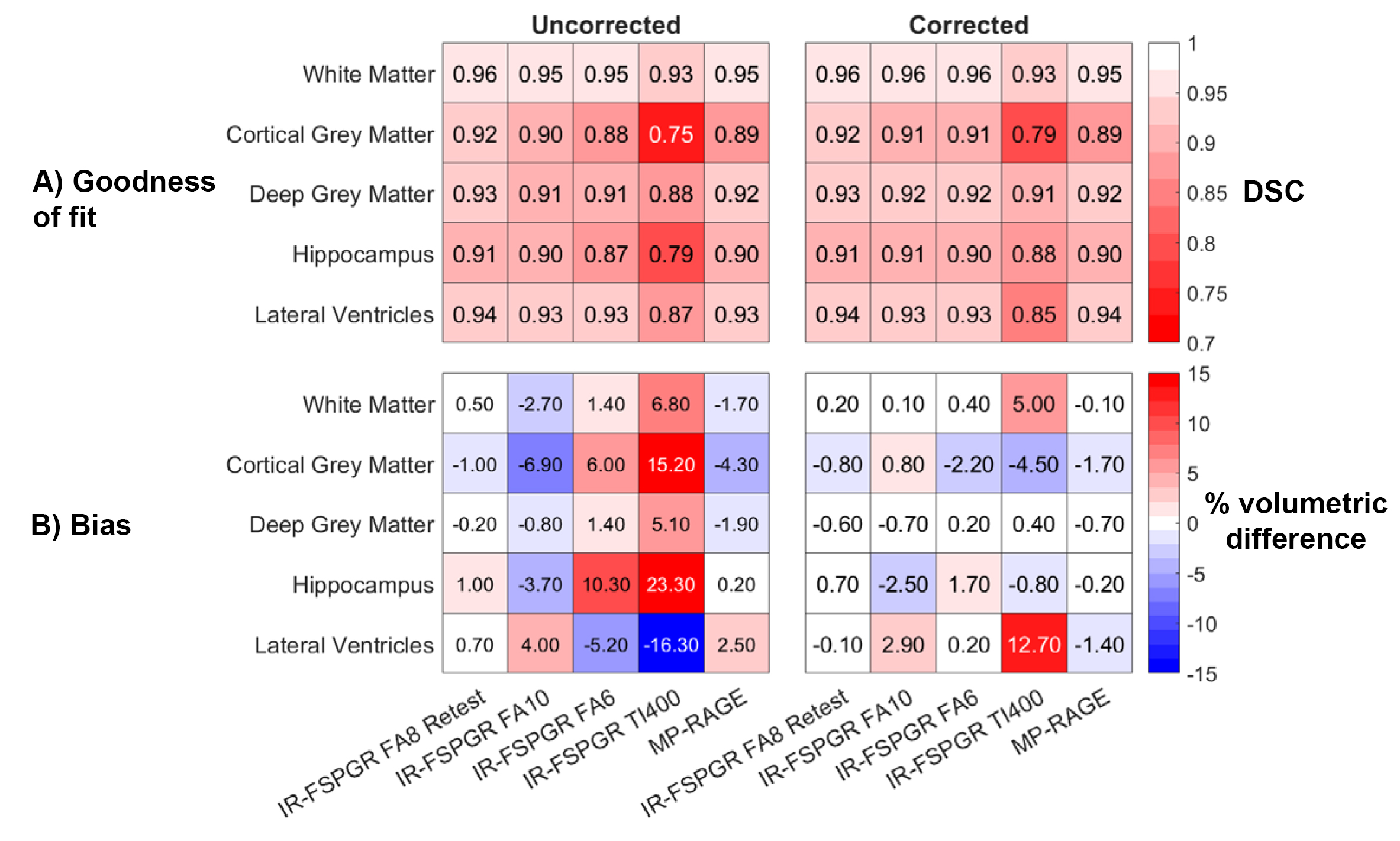
Figure 1: Goodness of fit and bias correction between target and source images before and after correction. Images were
corrected to the IR-FSPGR Flip Angle 8. A) The Dice Similarity Coefficient
(DSC) assesses the goodness of fit between each sequence. Higher values
(whiter) indicate better fit. B) Volumetric percentage difference between
target IR-FSPGR FA8 and each sequence.