2862
MR-only Radiation Therapy Planning workflow optimization for Head and Neck: Zero TE based pseudo CT conversion with body coil.1GE Healthcare, Munich, Germany, 2GE Global Research, Bangalore, India
Synopsis
Proton Density (PD) weighted Zero Echo Time (ZTE) imaging has been recently developed to provide bone, soft-tissue and air classification suitable for PET/MR Attenuation Correction and Radiation Therapy Planning (RTP). Here we demonstrate ZTE based derivation of pseudo CT using an optimized body coil protocol, which enables patient positioning in the MRI with the RT fixation devices, while preserving the image quality and reproducibility needed for pseudo CT conversion. The method was tested for the head and neck application on N=5 volunteers for different resolutions. The results were compared versus a high SNR surface coil, previously demonstrated suitable for pseudo CT conversion and dose calculation.
Introduction:
MR-only Radiation Therapy (RT) is an area of growing interest due to its soft tissue contrast and its suitability for Target Volume (TV) and healthy tissue delineation. Major issues currently being addressed are the pseudo CT conversion and the geometric accuracy of the chosen MR sequences. ZTE MR imaging was recently demonstrated suitable for both Attenuation Correction in PET/MR [1] and for pseudo CT conversion for Head and Neck applications [2]. Another important challenge in the MR-only workflow is the availability of appropriate coils. Surface coil are typically tuned for specific anatomies and may lead to sensitivity gaps dependent on placement and channel selection. Furthermore, they may be impractical or even incompatible with RT fixation devices. Body coil provides instead repeatable and uniform coil sensitivity and allows the use of RT fixation devices. These advantages however typically come at the known expense of a reduced SNR. The purpose of this study is to demonstrate ZTE based derivation of pseudo CT for MR-based RT planning using an optimized body coil protocol suitable for an MR-only workflow.Methods:
Five volunteers were enrolled in this study. Proton density weighted ZTE imaging was performed on a 3T GE MR750w scanner (GE Healthcare, Chicago, IL) using body coil receive and testing three different resolution protocols:
Resolution=2.4mm, acquisition time=60 sec, FA=1deg, BW=± 62.5kHz, FOV= 30.7 cm;
Resolution=2mm, acquisition time= 90 sec, FA=1deg, BW=± 62.5kHz, FOV= 30.7 cm;
Resolution=1.5mm, acquisition time= 190 sec, FA=1deg, BW=± 62.5kHz, FOV= 30.7 cm.
A GEM head and neck array coil (GE Healthcare, Chicago, IL) was used on one of the volunteer as reference.
ZTE data were processed with both a threshold-based pixel-wise conversion method [2] and with a Deep Learning (DL) method. For the DL method, a 3D convolutional neural network of the U-net architecture with 8-layers, Adam optimizer and RMSE cost function was adapted to perform pseudo-CT computation via image regression. This method was extensively trained on a diverse set of N=50 patients using matched pairs of ZTE and CT patient data sets [3]. A post-processing step was also applied to smooth soft tissue non-uniformities and a fixed value of 42HU was assigned.
Results:
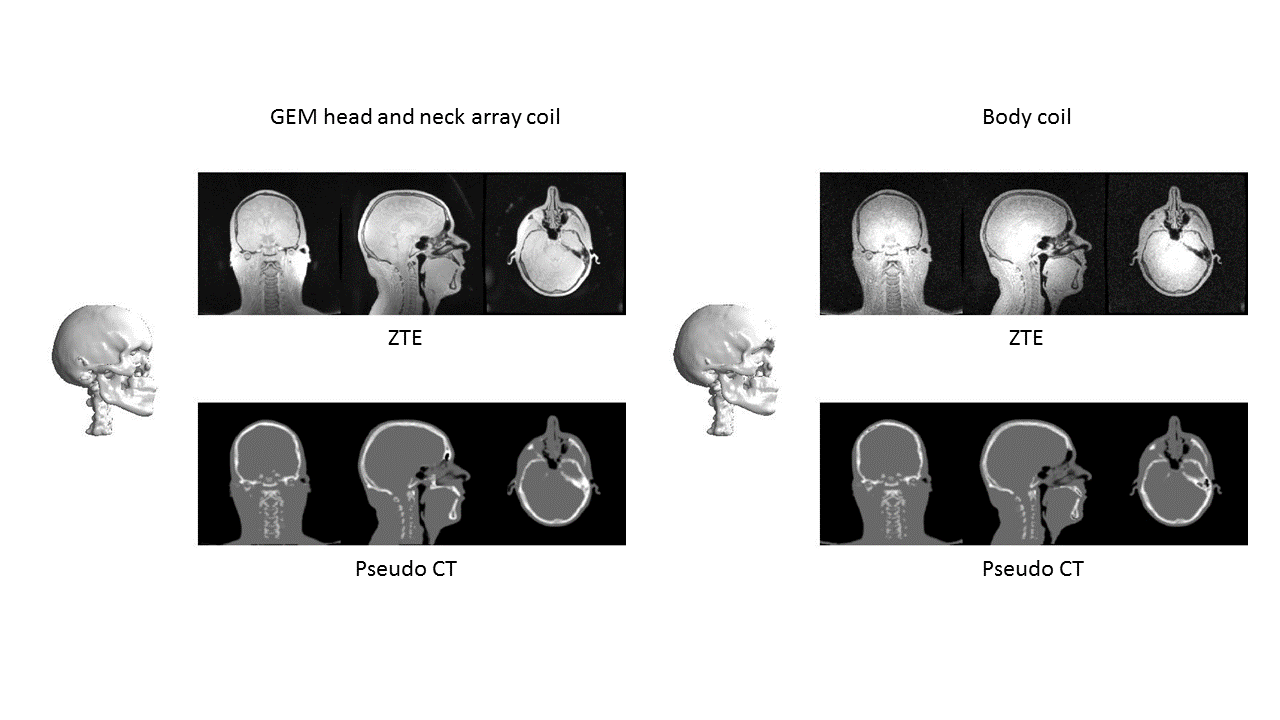
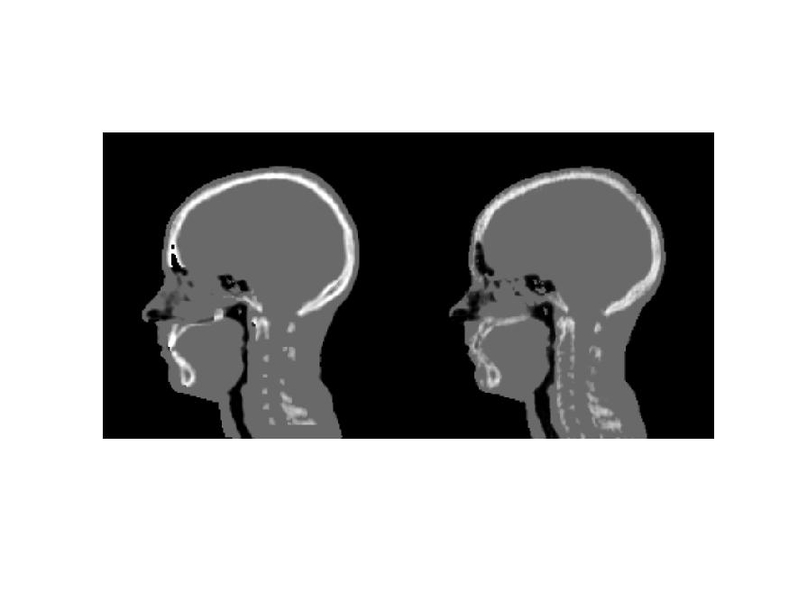
In Figure 1 the raw images acquired with the GEM head and neck array coil used as reference is shown in the left top row. On the right top row, the ZTE raw images from the body coil are shown. At the bottom, the pseudo CT converted data for the GEM head and neck array coil on the left and for the body coil on the right are illustrated. A 3D rendering of the bone is also shown for both coils. The body coil on the right provides uniform coverage (i.e. similar to the GEM head and neck array coil on the left) at the cost of reduced SNR which however does not significantly impact the pseudo CT result. To bridge the SNR gap, a DL method was used, which improves in particular the sinus depiction as shown in Figure 2. In Figure 3 the overview of the results from the pixel-wise conversion is shown. The top row shows the pseudo CT converted 2.4 mm isotropic resolution data and its corresponding bone mask; the middle row shows the results for 2 mm isotropic resolution while the bottom row illustrates the high-resolution results for all 5 volunteers.Conclusion:
We presented a body coil protocol which gives comparable results to the high SNR GEM head and neck array coil for pseudo CT conversion. We demonstrated the pseudo CT for different resolutions, including 1.5 mm isotropic resolution for both pixel-wise and DL conversion methods. These results indicate that the body coil can be used allowing for all required fixation devices for an optimized MR-only based RTP workflow. However, for normal anatomical imaging, surface array coils are still preferred because of their intrinsic SNR and speed advantage.Acknowledgements
No acknowledgement found.References
[1] F. Wiesinger et al., „Zero TE MR bone imaging in the head“, Magnetic Resonance in Medicine (2015)
[2] C. Cozzini et al., „Feasibility of Zero TE MR based Radiation Therapy Planning for Head application“, ISMRM 2017
[3] S. Kaushik et al., „Deep Learning based pseudo-CT computation and its application for PET/MR attenuation correction and MR-guided radiation therapy planning“, ISMRM 2018, submitted
Figures