2859
Generic feature extraction accompanied by support vector classification: an efficient and effective way for MR image quality determinationDirk Bequé1, Arathi Sreekumari2, Dattesh Shanbhag2, Keith Park3, Desmond Teck Beng Yeo3, Thomas K.F. Foo3, and Ileana Hancu3
1GE Global Research, Garching bei München, Germany, 2GE Global Research, Bangalore, India, 3GE Global Research, Niskayuna, NY, United States
Synopsis
Support vector machine image classification is performed on MR brain images to determine the need to repeat the MR acquisition. However, the image feature extraction is completely brain image agnostic. It is performed either directly on image slices or simple transformations thereof, like e.g. by fore/background thresholding or 1-level wavelet decomposition. 120 image features and meta-data entries are used to classify images as sufficient to diagnose or not. 84% accuracy is demonstrated, even after reducing the feature space to only 20 features. Such feature computation is fast enough to perform image quality assessment in real time, immediately after scan completion.
Introduction
Patient motion is a frequent cause of degraded image quality in MR imaging. Mild motion artifacts do not necessarily render an image clinically useless, yet severe artifacts require the acquisition to be repeated. Taking the decision to repeat or not requires considerable skill from the system operator. This study explores the possibility of making this decision in real time by generic image feature extraction and machine learning immediately after scan completion. The aim is to base the decision only on a limited set of generic image features that can be calculated very fast.Methods
Expert radiologists have classified the images of 200 neurological patient exams into acceptable and inacceptable image quality, with the acquisitions of the latter class needing to be repeated. Images with artifacts not caused by motion were excluded from the study. All acquisitions are 2D and performed at 1.5T, but include different orientations (axial, sagittal and coronal), various contrasts (including T1, T2, T2*, T1 FLAIR and T2 FLAIR) and both cartesian and propeller readouts. For the images of inacceptable quality, non-expert readers further identified the slices clearly affected by motion and discarded the other image slices. Finally, the images were randomly split into training and testing data for machine learning, resulting into the split of figure 1. For each labeled image slice, a total of 108 brain image agnostic features were calculated either on the entire slice, or simple decompositions thereof by e.g. fore/background thresholding, wavelet decomposition or discrete cosine transform. The features extracted included noise metrics, focus measures1, grey level co-occurrences and classical texture features2. These features were further supplemented with 12 meta data entries from the image header, including e.g. the repetition and echo time, the flip angle and the slice orientation. At first a support vector machine (2 class C-SVM with radial basis function kernel, LibSVM 3.223) was trained to classify slices of acceptable and inacceptable image quality based on the full set of 120 features. The optimal SVM hyperparameter values were determined by grid search and cross-validation with a leave (all slices of) 15 series out strategy. To partially offset the large imbalance between the number of slices with acceptable and inacceptable quality, the inacceptable slices were weighted higher accordingly, yet without attempt to optimize this weight. Next, recursive feature elimination4, eliminating just a single feature at a time, was performed to the 120 features to rank them according to their importance for the classification. Afterwards, a new support vector training was performed using only the 20 top-ranking features. Finally, the performance of the 2 SVM’s is compared.Results
The 120 feature SVM yielded an overall accuracy of classification of 90.1% on the validation data and 85.7% on the testing data. Figure 2 provides a complete overview of the performance. The 20 feature SVM yielded only slightly worse performance with an overall classification accuracy of 87.7% on the validation data and 83.3% on the testing data (see also figure 2). For both SVM’s, the classification performance on acceptable slices is further somewhat higher than for inacceptable slices. From the 120 features, the recursive feature elimination selected 2 meta data features: the repetition time and the flip angle of the sequence.Discussion
The decision of whether an MRI acquisition is of sufficient quality for diagnosis can be difficult to make. This study has investigated the possibility to automate/facilitate this decision in real time by generic image feature extraction and machine learning. The study was limited to brain imaging, but included a wide range of different protocols of clinical interest. Furthermore, the features used do not require any brain specific image processing like e.g. gray/white matter segmentation. This ensures fast and reliable feature extraction and it also makes the method easily applicable to other body parts and protocols. The results of the study are encouraging, especially when taking into account that the results provided are for the classification of individual slices. In practice, a decision for an entire series volume is required based on the classifications of the composing slices. The misclassification of just a few slices doesn’t have to lead to the misclassification of the entire volume. The study has also demonstrated that, unlike in other studies5, a limited set of features can already be sufficient for good performance. The results finally also suggest that the inclusion of specific sequence parameters is beneficial for the classification accuracy.Conclusion
Generic image feature extraction in combination with support vector machine learning is a promising method to judge the clinical quality of MR images.Acknowledgements
No acknowledgement found.References
- Pertuz S et al. Analysis of focus measure operators for shape-from-focus. Pattern Recognition, 46(5), 1415-1432, 2013.
- Haralick RM, Shanmugan K, and Dinstein I. Textural Features for Image Classification. IEEE Trans. On Systems, Man and Cybernetics, Vol 3(6), pp. 610-621, 1973.
- Chang C-C and Lin C-J. LIBSVM : a library for support vector machines. ACM Transactions on Intelligent Systems and Technology, 2:27:1--27:27, 2011.
- Guyon I, Weston J, et al. Gene Selection for Cancer Classification using Support Vector Machines. Machine Learning, 46, 389–422, 2002.
- Gatidis S, Liebgott A, et al. Automated reference-free assessment of MR image quality using an active learning approach: Comparison of Support Vector Machine versus Deep Neural Network classification. ISMRM 2017.
Figures
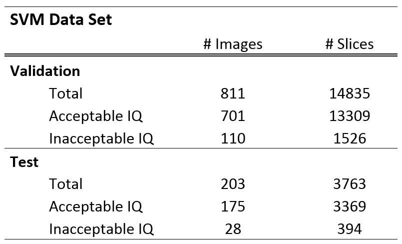
Figure 1: Support
vector classification data set.
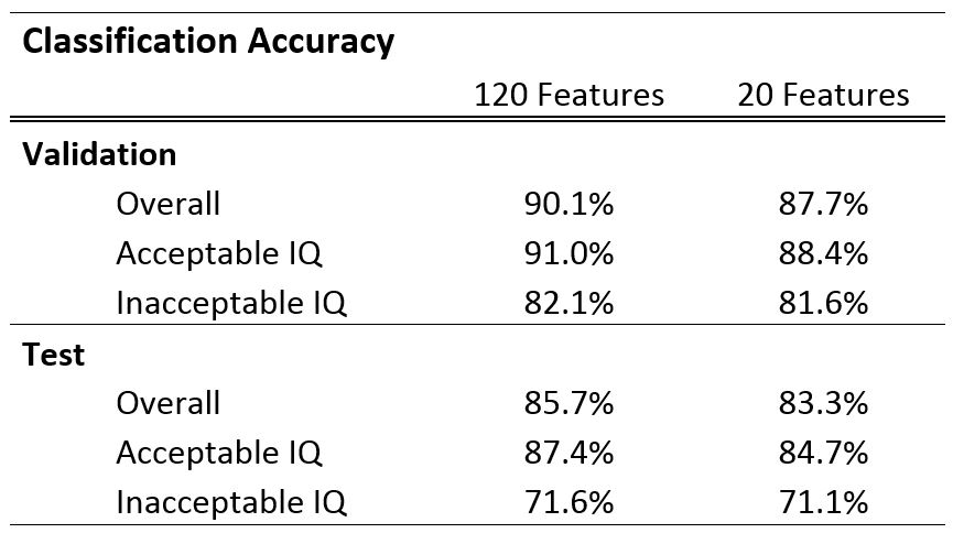
Figure 2: Overview
of the classification accuracy of the SVM’s.