2848
A Simplified Framework for MR Image Processing & 3D Printing in Healthcare Applications1Centre for Biomedical Engineering, Indian Institute of Technology Delhi, New Delhi, India, 2Department of Electronics & Communication Engineering, G L Bajaj Institute of Technology and Management, Greater Noida, India, 3Department of Electrical Engineering, Indian Institute of Technology, New Delhi, India, 4Department of Chemical Engineering, Indian Institute of Technology, New Delhi, India, 5Department of Radiology, Medanta: The Medicity Hospital, Gurugram, India
Synopsis
This work summarized a simplified framework that can be used to generate 3D printed prototype from 3D MRI images, with the help of widely available processing tools. The process is conceptually divided into three steps: image acquisition, image post-processing and 3D printing. The utility of the streamlined framework is demonstrated by building 3D prototype of Liver, Spleen and Kidneys using Selective Laser Sintering (SLS) and Fused Deposition Modeling (FDM) technology based 3D printers. The simplified approach has been suggested to assist users in creating 3D anatomical model from medical imaging data using relevant open source tools.
Introduction:
3D reformatting in MR imaging along with 3D printing innovation is improving our insight of disease diagnosis and treatment planning in a better way. It has discovered a significant impact in biomedical field by creating patient-specific implants and sensible anatomical models for surgical–interventional training and planning. There are multiple approaches for 3D printed models that can be generated from 3D MRI [1], users may be lost in this complex process, therefore a streamlined pipeline is essential. This abstract present the implementation of a framework using various examples to represent the simplified approache that can be followed.Methods:
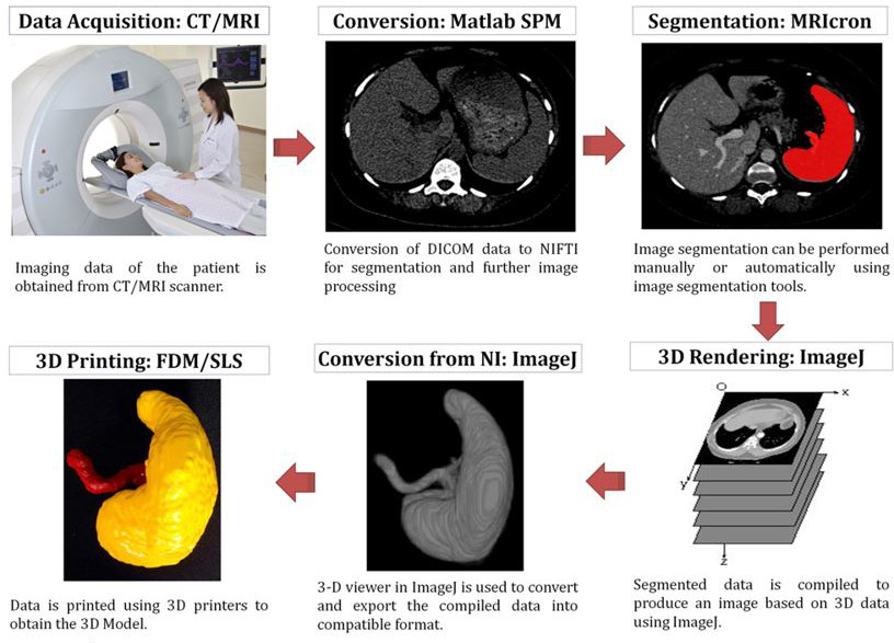
For creating 3D printed model, required anatomy is defined by marking the region of interest over acquired MR images by an expert [2]. Further, Image post-processing algorithm are used for 3D rendering and smoothing the desired model. These structures need to convert to compatible STL format for 3D printing [1], [3]. This complex process can be conceptually divided into three simple steps: i) image acquisition, ii) image post-processing and iii) 3D printing [4], [5]. The Figure 1 show these steps involved from image acquisition to 3D printing.
Data acquisition: DICOM: Digital Imaging and Communications in Medicine (.dcm), is the standard format in which the medical imaging data is acquired from the MRI scanner [6]. DICOM images can be converted to NIfTI: Neuroimaging Informatics Technology Initiative (.nii) format this can be performed using Matlab SPM toolbox for simplified post-processing [7].
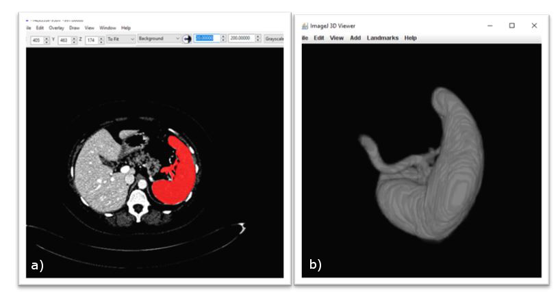
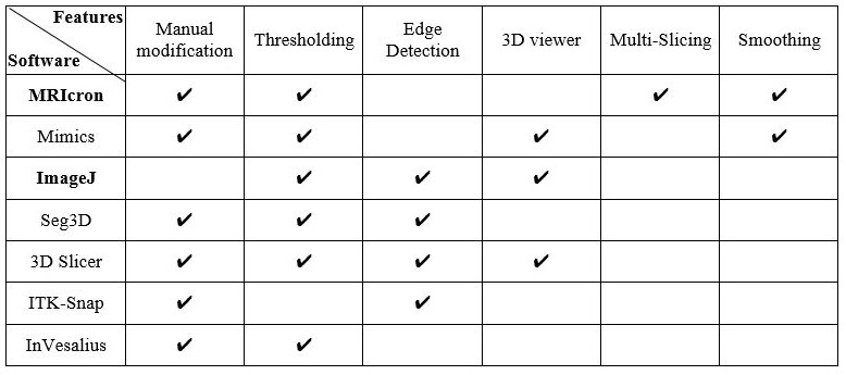
Image post processing: The dedicated 3D post-processing tools can be used for working on NIfTI images [8]. The post-processing generally includes segmentation, volume/surface rendering, smoothening and multiplanar reformation [1], [9]. Figure 2 display the MRIcron software used for marking ROI mask and the 3D model of Spleen generated by 3D viewer plugin of ImageJ freeware. 3D viewer plug-in of ImageJ can be utilized to transform segmented mask, NIfTI(.nii), into STL: Stereolithography (.stl) 3D model [22]. Stereolithography is a standard format for 3D printing, widely accepted across various 3D printing technologies and vendors. Table 1 present advantaged/limitation of various other tools that can be used instead.
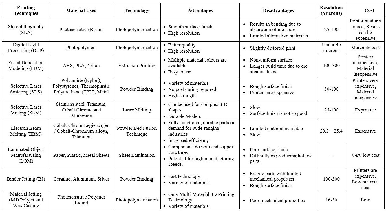
3D printing: After getting a virtual 3D model (.stl/.vtk) of particular anatomy of interest, the next important step involves selecting a suitable technology/material for the 3D model. Table 2 elaborates the various 3D printing technologies with their specifications, advantages and limitations [6]. It is important to understand the technology and material suitable for the required specific clinical application. The smoothing of 3D rendered model could be essential before moving forward. This can be obtained by process the stack of segmented slices by open source visualization toolkit (VTK) using Matlab [10].
Results:
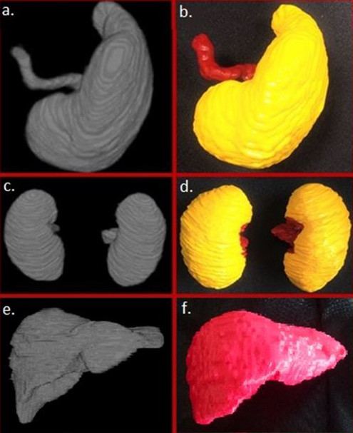
A simplified framework has been introduced that can be utilized to create 3D printed models from volumetric MRI data. Using this generalized workflow, 3D printed models of the spleen, kidneys and liver has been demonstrated in Figure 3. Here, spleen and kidneys are printed with Selective Laser Sintering (SLS) technology using PA (Poly Amide) material while liver is printed with Fused Deposition Modeling (FDM) technique using PLA material.Discussion:
The basic step for creating 3D models is to segment the medical images. There are various open source tools that are available as discussed in Table 1. The MRIcron is one of the freeware for manual image segmentation, which allows user to segment the organ of interest from NIfTI images using multi slicing segmentation [12]. The segmented model (DICOM/NIfTI) could be transformed into virtual 3D model (.stl) using various CAD software or tools [13]; one easy solution is 3D viewer plug-in of ImageJ [14]. Furthermore, it is possible to build detailed anatomical structure using several smoothing techniques [5]. One common problem among all 3D printing technologies is that one may end up losing several vital details of the anatomy if an attempt is made to smoothen the surface beyond an optimal limit [11]. Similarly, different 3D printing technologies are also available for printing the 3D model [1]. Fused Deposition Modeling is the commonly used and cost effective 3D printing technology [13]. However, Selective Laser Sintering gives better print quality with higher cost.Conclusion:
The abstract demonstrated a simple and easy to implement process pipeline for 3D printing of patient specific MRI using open source tools. This presented framework can be used for medical education and training but have significant potential for development of new healthcare applications. The framework would be shared with user on laptop and hands-on session can be delivered at the abstract presentation site.Acknowledgements
Authors would like to thank IIT Delhi for funding support under IRD project DL1-2-3-4, project number MI01511.References
[1] T. M. Bücking, E. R. Hill, J. L. Robertson, E. Maneas, A. A. Plumb, and D. I. Nikitichev, “From medical imaging data to 3D printed anatomical models,” PLoS One, vol. 12, no. 5, pp. 1–10, 2017. [2] N. Sharma and L. M. Aggarwal, “Automated medical image segmentation techniques.,” J. Med. Phys., vol. 35, no. 1, pp. 3–14, Jan. 2010. [3] J. . Taboas, R. . Maddox, P. . Krebsbach, and S. . Hollister, “Indirect solid free form fabrication of local and global porous, biomimetic and composite 3D polymer-ceramic scaffolds,” Biomaterials, vol. 24, no. 1, pp. 181–194, Jan. 2003. [4] F. Rengier et al., “3D printing based on imaging data: Review of medical applications,” Int. J. Comput. Assist. Radiol. Surg., vol. 5, no. 4, pp. 335–341, 2010. [5] C. N. Ionita et al., “Challenges and limitations of patient-specific vascular phantom fabrication using 3D Polyjet printing.,” Proc. SPIE--the Int. Soc. Opt. Eng., vol. 9038, p. 90380M, Mar. 2014. [6] M. Larobina and L. Murino, “Medical Image File Formats,” J. Digit. Imaging, vol. 27, no. 2, pp. 200–206, Apr. 2014. [7] G. Queirós, “Computational Methods for fMRI image Processing and Analysis,” 2013. [8] X. Li, P. S. Morgan, J. Ashburner, J. Smith, and C. Rorden, “The first step for neuroimaging data analysis: DICOM to NIfTI conversion,” J. Neurosci. Methods, vol. 264, pp. 47–56, 2016. [9] T. L. Sanders, “Image Processing and 3-D Reconstruction in Tomography,” 2010. [10] W. Schroeder, K. Martin, B. Lorensen, L. S. Avila, R. Avila, and C. Law, “The Visualization Toolkit An Object-Oriented Approach To 3D Graphics Third Edition.” [11] N. Green, V. Glatt, K. Tetsworth, L. J. Wilson, and C. A. Grant, “A Practical Guide to Image Processing in the Creation of 3D Models for Orthopedics,” Tech. Orthop., vol. 31, no. 3, pp. 153–163, Sep. 2016. [12] D. L. Pham, C. Xu, and J. L. Prince, “Current Methods in Medical Image Segmentation,” Annu. Rev. Biomed. Eng., vol. 2, no. 1, pp. 315–337, Aug. 2000. [13] C. L. Ventola, “Medical Applications for 3D Printing: Current and Projected Uses.,” P T, vol. 39, no. 10, pp. 704–11, Oct. 2014. [14] M. D. Abràmoff, P. J. Magalhães, and S. J. Ram, “Image Processing with ImageJ.”Figures