2834
Beyond high resolution: Pitfalls in quantification of cortical thickness based on higher and ultra-high resolution data1Biomedical Magnetic Resonance, Otto-von-Guericke University, Magdeburg, Germany, 2Center for Behavioral Brain Sciences, Magdeburg, Germany, 3Leibnitz Institute for Neurobiology, Magdeburg, Germany, 4German Center for Neurodegenerative Disease (DZNE), Magdeburg, Germany
Synopsis
It was shown that higher resolution data increases the accuracy of the brain segmentation resulting in a decrease of cortical thickness estimates. However, data is still mostly acquired at a spatial resolution of 1 mm for quantifying cortical thickness. Several software packages allow processing of higher resolution data. However, FreeSurfer constitutes the de facto standard due to its prevalence. Therefore, we investigate the effects of resolution and SNR at two important stages of its standard processing pipeline: the skull stripping and white matter segmentation.
Introduction
It was previously shown, that the accuracy of the segmentation increases while cortical thickness estimates decrease with higher spatial resolution1-3. However, an isotropic spatial resolution of 1mm still is considered the clinical and neuroscientific standard for quantification of cortical thickness. Even at 7T the resolution is scarcely higher than 0.7mm. Why is that so? Is it the decrease in signal-to-noise ratio (SNR) in higher resolution acquisitions which is not entirely compensated by higher field strength? Or is it the longer scan time that inherently introduces more motion artifacts in higher resolution acquisitions? At least the latter has been recently tackled with the establishment of retro- and prospective motion correction methods that can remediate subject motion4-7.
FreeSurfer constitutes the de facto standard in determining cortical thickness due to its prevalence, but other software is capable of processing higher resolution data also (e.g., CBS-Tools, ANTs, and CAT12). Within the standard processing pipeline of FreeSurfer are two important intermediate stages: the skull stripping and white matter segmentation. The importance of the skull stripping procedure is obvious as any remaining structures besides the brain may be a potential confound in segmentation. The importance of the white matter segmentation is less obvious if cortical grey matter is to be quantified. Based on the volumetric white matter segmentation the white matter surface is generated. The white matter surface is then inflated until an abrupt intensity drop is observed – the boundary between cortex and cerebrospinal fluid. Here, we investigate potential pitfalls at these two stages of FreeSurfer’s processing pipeline related to resolution and SNR.
Methods and Materials
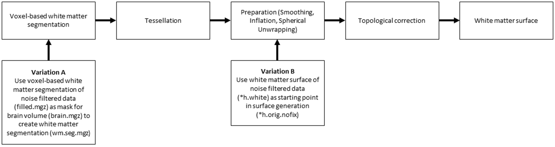
We have acquired T1-weighted MPRAGE data with an isotropic spatial resolution of 0.5mm of a single subject with prospective motion correction [9] and TR-FOCI inversion pulse10. Data were acquired with higher than usual bandwidth (200Hz/px instead of 130Hz/px) without any acceleration to produce a fairly low SNR volume without any potential acceleration related artifacts. The data are bias field corrected with SPM12 with parameters for 7T data. Then they are processed with FreeSurfer using the ‘hires’ flag to keep the native resolution and without it to resample the data to 1mm. Additionally, the data were denoised with the BM4D11 algorithm and also processed with the ‘hires’ flag. Furthermore, the voxel-based white matter segmentation and the resulting white matter surface segmentation of the denoised data have been used in the unfiltered processing stream to evaluate the effects in two variations (Fig.1).Results
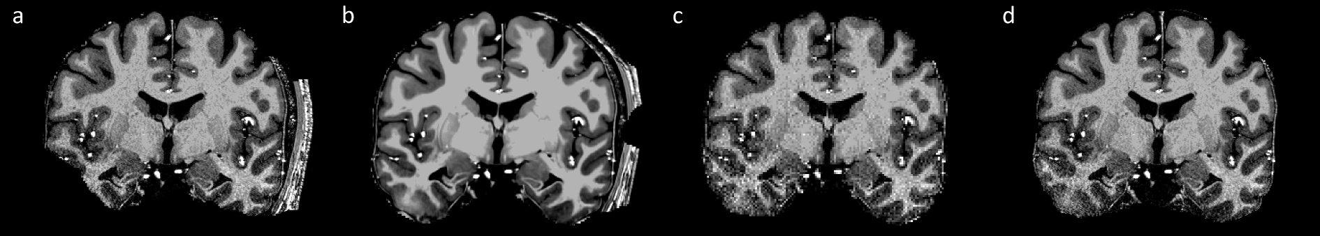
Skull stripping is insufficient if native unfiltered data are used (Fig.2,a). Denoising does not help in getting rid of the remaining scalp (Fig.2,b). Skull stripping is much improved if data are resampled to 1 mm (Fig.2,c). As a byproduct of SPM’s bias field correction segmentations of WM, GM and CSF are provided. The volume generated by adding the segmentations into a single volume can be used to mask the brain of the pre-processed FreeSurfer data. This results in a very accurately skull-stripped brain (Fig.2,d).
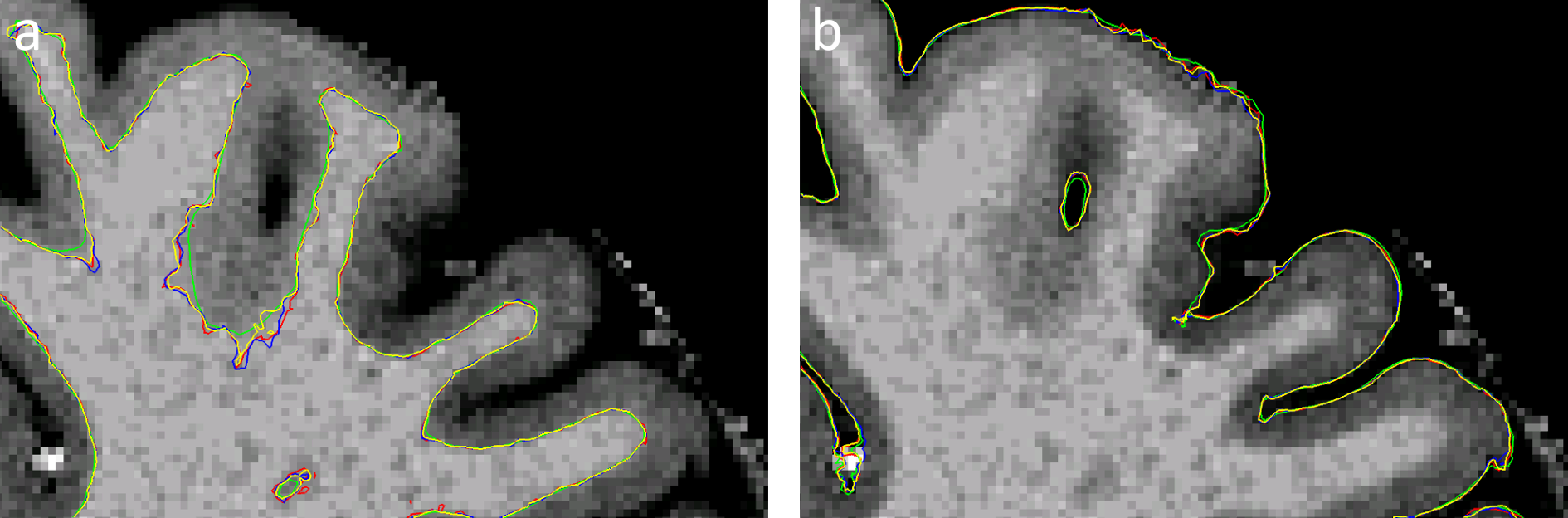
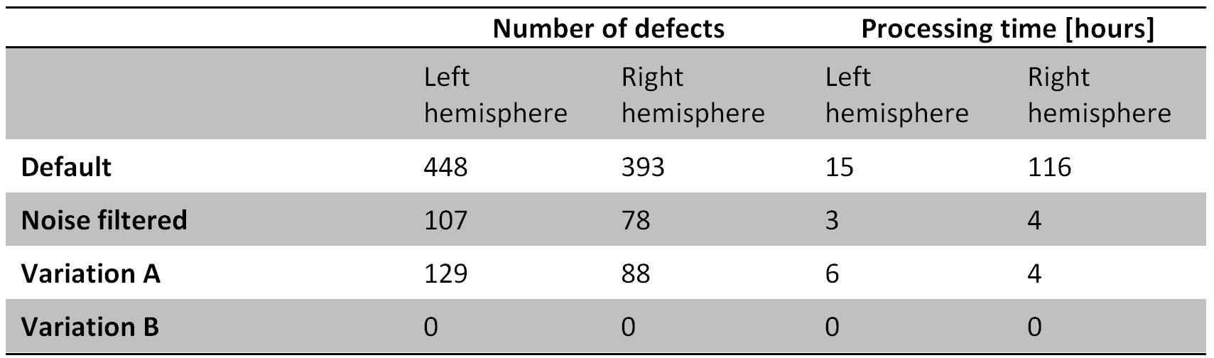
Denoising the data causes a displacement of the white surface (Fig.3,a). The white matter surface generated by ‘Variation A’ and ‘Variation B’ qualitatively follows the surface of the ‘hires’ processing stream (Fig.3,a). However, while surface placement does not deviate much from the default processing stream, the processing time to generate the surfaces is reduced tremendously (Tab.4). The gray matter surface generation is neither affected by denoising nor in ‘Variation A’ or ‘B’ (Fig.3,b).
Conclusion
The skull stripping procedure of FreeSurfer is not sufficiently capable of handling higher resolution data. This can be overcome by using a brainmask, either generated by resampling the data to 1mm [ref. 1] or by using the byproduct of SPM’s bias field correction approach. As the bias field correction of SPM outperforms FreeSurfer’s bias field correction11 the latter approach is recommended. Denoising reduces the processing time of the topological correction and affects surface positioning, presumably overcorrecting it. By using either presented variations the white matter surface follows the segmentation of the unfiltered data stream more closely. However, it remains unclear whether either approach results in a more accurate segmentation. This has to be evaluated in a future study by adapting the approach of Fujimoto et al.12 to investigate the surface positioning on a vertex-basis as has been recently done3. Nonetheless, the processing time of both variations is tremendously reduced and may be a viable option in processing higher or ultra-high resolution data in the future.
Acknowledgements
No acknowledgement found.References
[1] Lüsebrink et al (2013) “Cortical Thickness determination of the human brain using high resolution 3T and 7T MRI data.” NeuroImage
[2] Glasser el al (2013) “The minimal processing pipelines for the Human Connectome Project.” NeuroImage
[3] Zaretskaya et al (2017) “Advantages of cortical surface reconstruction using submillimeter 7T MEMPRAGE.” NeuroImage
[4] Stucht et al (2015) “Highest Resolution In Vivo Human Brain MRI Using Prospective Motion Correction” PlosONE
[5] Gallichan et al (2016) “Motion-Correction Enabled Ultra-High Resolution In-Vivo 7T-MRI of the Brain” PlosOne
[6] Watanabe et al. (2016) “Utility of real-time prospective motion correction (PROMO) on 3D T1-weighted imaging in automated brain structure measurements” Sci. reports
[7] Lüsebrink et al. (2017) “T1-weighred in vivo human brain whole brain MRI dataset with an ultrahigh isotropic resolution of 250 µm” Sci. data
[8] Maclaren et al. (2013) “Measurement and correction of microscopic head motion during magnetic resonance imaging of the brain.” PlosONE
[9] Hurley et al. (2010) “Tailored RF Pulse for Magnetization Inversion at Ultrahigh Field” MRM
[10] Maggioni et al. (2012) “Nonlocal Transform-Domain Denoising of Volumetric Data With Groupwise Adaptive Variance Estimation” Proc. SPIE Electronic Imaging
[11] Lüsebrink et al. (2017) “Quantitative and qualitative evaluation of bias field correction methods” ISMRM 2017 #1451
[12] Fujimoto et al. (2014) “Quantitative comparison of cortical surface reconstructions from MP2RAGE and Multi-Echo MPRAGE data at 3 and 7 Tesla” NeuroImage
Figures