2827
Laplacian pyramid based data fusion for high resolution dynamic MRILiad Pollak Zuckerman1, Lior Weizman2, Yonina C. Eldar2, Dafna Ben Bashat3, Moran Arzi3, and Michal Irani1
1Faculty of Mathematics and Computer Science, Weizmann Institute of Science, Rehovot, Israel, 2Department of Electrical Engineering, Technion - Israel Institute of Technology, Haifa, Israel, 3Tel Aviv Medical Center, Tel Aviv University, Tel Aviv, Israel
Synopsis
Dynamic contrast-enhanced (DCE) MRI is useful for tumor diagnosis and treatment. In DCE, there is a tradeoff between the spatial and temporal resolutions. Improving the spatial resolution while preserving the temporal dynamics is essential for better diagnosis/treatment. We present a method (LAPFUD) for enhancing the spatial frequency without compromising on temporal resolution. LAPFUD combines information from a static high-resolution image acquired at baseline, with each low-resolution frame. By making local decisions it preserves details from both inputs without changing the temporal behavior. Experiments show that LAPFUD provides superior performance (spatially and temporally) compared to the commonly used keyhole method.
Introduction:
Dynamic contrast-enhanced (DCE) magnetic resonance imaging has been widely used to characterize the microvasculature of brain lesion and tumors1,2. High temporal resolution (< 2 sec)3 is only required in the first minute post contrast agent injection, for accurate detection of bolus arrival time, while high spatial resolution is necessary for diagnosis. There is an inherent tradeoff between temporal and spatial resolutions, dictated by the physics of the sequence used, affecting also brain coverage. Several methods have been proposed to enhance the spatial resolution of the dynamic images based on high-resolution (HR) images acquired in the first minute post injection4, or to enhance the temporal resolution based on partial acquisition of the data5. The keyhole method6, which is implemented in many clinical DCE sequences, is based on the acquisition of static HR images before the dynamic sequence. It combines, in the k-space domain, high-frequency data taken from the HR images, with low frequency data acquired during the dynamic acquisition. The aim of this study is to propose a new method, to improve the spatial resolution of dynamic images based on an HR image acquired at baseline. The proposed method coined LAplacian Pyramid based FUsion for improved DCE (LAPFUD) fuses between the HR image and the low-resolution (LR) dynamic sequence using Laplacian pyramids7. Compared to the keyhole approach that acts globally on the entire image, LAPFUD makes local decisions on selecting the strongest local edges from either the LR or HR images. Therefore, it captures details from both inputs (the HR image and LR sequence) and results in many spatial details that do not exist in the keyhole method, without compromising on the temporal resolution of the original LR sequence.Methods:
A static HR MR image was acquired prior to dynamic acquisition. Both the HR image and the LR sequence consisted of 20 axial slices, with matrix size of 512x368 for the HR image and 256x184 for the LR DCE sequence, which contained a total of 78 LR frames acquired with temporal resolution of 6 seconds. Each slice in the LR frame is interpolated (bicubic interpolation) by 2 to match the HR image size. Then, realignment and reslicing was performed using the SPM tool8. For both HR and LR frames a Laplacian pyramid9 was calculated with depth 4. Each pyramid level captures information about a different spatial frequency range, except the lowest level which is a small down-scaled version of the original. We then form a new Laplacian pyramid by choosing, at each pixel, the coefficient from the transformed HR and LR images with the highest absolute value7. Effectively, LAPFUD corresponds to selecting the strongest local edges at each frequency range. The final fused image is obtained by an inverse transform on the result. The LAPFUD algorithm is illustrated in Figure 1. For comparison purposes, the keyhole method is also applied on the same HR and LR images.Results and Discussion:
Figure 2 shows data obtained from patient with high grade brain tumor: The input data (slice 11 taken from both the HR and LR images) and the results of both keyhole and our proposed LAPFUD. The contrast agent enhancement is clearly visible in the LR image (frame #13 during contrast agent injection). By applying LAPFUD, more detailed information from the HR image is preserved. To ensure that the temporal dynamics has not been affected by LAPFUD, we examined two pixels' intensity profiles over time, taken from homogenous healthy and tumor regions. The time courses of the original LR sequence, the keyhole result and LAPFUD for a healthy and tumor pixel are shown in Figure 3. Both keyhole and LAPFUD exhibit no major changes compared to the original LR sequence (both provide temporal correlation with the original LR time course of above 0.99) for the 2 pixels examined. In other words, LAPFUD exhibits improved spatial resolution (compared to keyhole) while preserving the temporal one.Conclusion:
We presented a method to enhance the spatial resolution in DCE MRI by using a HR static image. The Laplacian pyramid based fusion allows making local decisions and combining detailed information from both images. We illustrated that this method improves spatial resolution while preserving temporal dynamics, leading to improved temporal/spatial resolution tradeoff. As a result, it can be used in cases where both high temporal and spatial resolutions of the dynamic sequence are required, for better diagnosis and treatment decisions making. Future work will examine LAPFUD performance in improving spatial resolution for DCE acquired at higher frame rates.Acknowledgements
No acknowledgement found.References
1P.Tofts ed. Quantitative MRI of the brain: measuring changes caused by disease. John Wiley & Sons, 2005;2Khalifa F, Soliman A, El-Baz A, Abou El-Ghar M, El-Diasty T, Gimel'farb G, Ouseph R, Dwyer AC. Models and methods for analyzing DCE-MRI: A review. Medical physics. 2014 Dec 1;41(12).
3Nadav, G., Liberman, G., Artzi, M., Kiryati, N. and Bashat D.B., 2017. Optimization of two-compartment-exchange-model analysis for dynamic contrast-enhanced mri incorporating bolus arrival time. Journal of Magnetic Resonance Imaging, 45(1), pp.237-249.
4I.O Jelescu et al. Dual-temporal resolution dynamic contrast-enhanced MRI protocol for blood-brain barrier permeability measurement in enhancing multiple sclerosis lesions. Journal of Magnetic Resonance Imaging May 2011.
5Otazp R, Candes E, Sodickson DK. Low-rank plus sparse matrix decomposition for accelerated dynamic MRI with separation of background and dynamic components. Magnetic Resonance in Medicine. 2015 Mar 1;73(3):1125-36
6Vanvalls JJ, et al. JMRI 1993 3(4):671-675.
7P. J. Burt, et al, Enhanced image capture through fusion in Proc. 4th Int. Conf. Computer Vision, Berlin, Germany, May 1993, pp 173-182.
8K. Friston et al. Statistical parametric mapping (SPM), 1999.
9P. J. Burt, et al, The Laplacian Pyramid as a Compact Image Code. IEEE transactions on communications, vol. com-31 no. 4, April 1983.
Figures
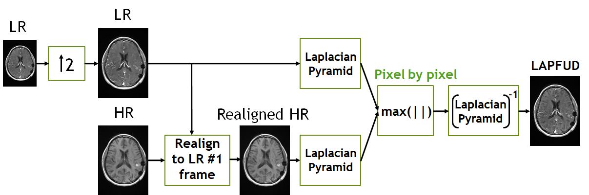
Figure
1: LAPFUD block diagram. First the LR
image is interpolated. Then the HR image and LR sequence are realigned and
resliced to match the first LR frame. Laplacian pyramids are calculated, and
the output image is reconstructed from the max absolute value from each pixel
in each level. The pixel with max absolute value is more informative as edges are
represented by high intensity pyramid values.
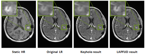
Figure 2: Representative result. The
two leftmost images are the input HR and LR images for slice 11. The two
rightmost images are the resulting outputs for the keyhole method and LAPFUD.
The green squares demonstrate a region of interest where details are better
preserved when using LAPFUD. The red
squares further show that due to its local nature, LAPFUD preserves local
spatial details better than the keyhole method.
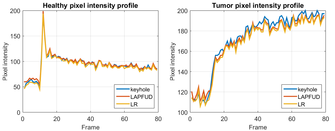
Figure
3: Pixel intensity profiles over time. Left - healthy pixel.
Right – tumor pixel. Blue – keyhole reconstruction. Red - LAPFUD
reconstruction. Yellow – LR sequence (original). In both methods the temporal
behavior is preserved (LAPFUD preserves the temporal behavior even slightly
better).