2818
Combination of Narrow-band KWIC and GROWL for Multiple T1-weighted Images Reconstruction Based on 3D Golden Angle Radial MR Sequence1Center for Biomedical Imaging Research, School of Medicine, Tsinghua University, Beijing, China, 2Neusoft Medical System, Shanghai, China
Synopsis
Radial sampling has been an increased application due to its insensitivity to motion. A reconstruction method combined the 3D GRAPPA operator for wider radial bands (GROWL) and narrow-band k-space weighted image contrast (KWIC) was proposed and used in GOAL-SNAP sequence. The proposed reconstruction method showed lower image RMSE, accurate T1 map estimation in simulation and higher image quality in in-vivo experiments with shorter computation time.
Introduction
Radial sampling has been an increased application due to its insensitivity to motion compared to Cartesian sampling1. Recently, a sequence consisting of an inversion recovery prepared pulse and a series of 3D golden angle radial acquisitions called GOAL-SNAP2 was proposed. The sequence had the ability to reconstruct multiple T1-weighted images using narrow-band k-space weighted image contrast (KWIC) algorithm and obtain T1 map from a single k-space dataset, thus was applied to detect T1 changes in carotid atherosclerotic plaques2. The use of narrow-band KWIC could improve image contrast but lead to undersampled k-space data3. In this study, a reconstruction method combined the 3D GRAPPA operator for wider radial bands (GROWL) and narrow-band KWIC was proposed to reduce aliasing artifacts, improve image quality and the accuracy of T1 estimation in 3D radial reconstruction.Methods
Theory: First, each 3D golden angle radial projection was expanded to two parallel projections along the azimuthal direction and two parallel projections along the zenithal direction using 3D GROWL method4, the GRAPPA operator was calculated using the fully sampled center k-space data. Then, 3D narrow-band KWIC method3 was applied to both the original and expanded projections paired. The width of the narrow band was determined by the temporal width (TW). Last, density compensation5 and gridding reconstruction were performed.
Simulation: 9 cylinders with T1 ranging from 200ms to 1800ms (interval=200 ms) were used in the simulation. GOAL-SNAP sequence (FOV=100*100*100 mm3 (2-fold oversampling); voxel size=0.8*0.8*0.8 mm3; TR/TE=11.3/4.7ms; flip angle=8o; TFE factor=155; IRTR=2000ms) as described in (2) was used to generate the simulated 3D k-space data. A total of 23250 projections were generated in the k-space to mimic a 5-minute scan. 1% Gaussian noise was added to both the real and imaginary parts of the simulated k-space.
In-vivo experiment: A patient with carotid atherosclerotic plaques was scanned on a 3T Philips Scanner using an 8-channel head coil with the same parameters as the simulation.
Reconstruction and analysis: 3D narrow-KWIC6, narrow-KWIC with CG-SENSE3, and the proposed narrow-KWIC with GROWL reconstruction methods were applied respectively to generate multiple T1-weighted images with TW=11, 15, 19, 23, 27 on the simulated k-space data and TW=19 on the in-vivo k-space data. The reconstruction time of the simulated and in-vivo data was recorded. Root mean square error (RMSE) was calculated between the reconstructed images and the reference images in the simulation. Signal intensity versus time curve was extracted voxel by voxel and fitted to the theoretically calculated signal equation7 to generate T1 maps. RMSE of the T1 maps estimated using three reconstructed methods were calculated in the simulation.
Results
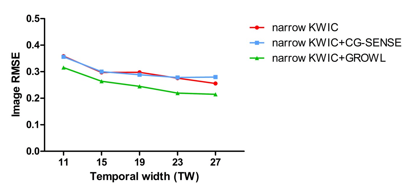
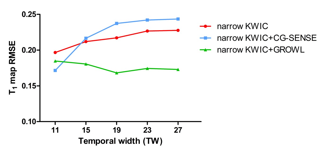
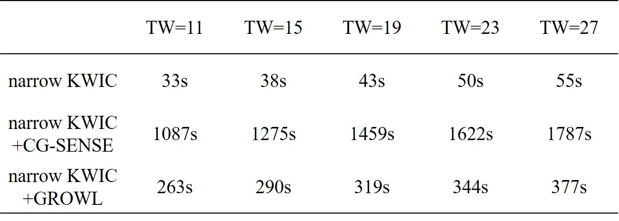
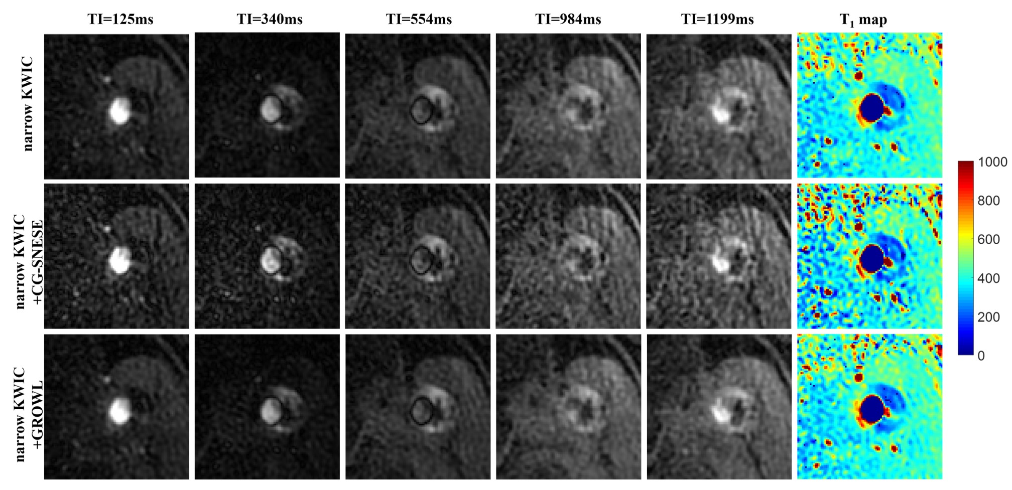
The narrow KWIC+GROWL showed lower image RMSE (Fig. 1) and lower T1 map RMSE (Fig. 2) than narrow KWIC and narrow KWIC+CG-SENSE. The computation time of simulation datasets at first TI using narrow KWIC+GROWL was approximately 3 times faster than narrow KWIC+CG-SENSE (Table. 1) . In-vivo images and estimated T1 maps with TW=19 were shown in Fig. 4. Narrow KWIC+GROWL showed better image quality and lower variations in T1 maps than the other two methods. The computation time of the in-vivo datasets at the first TI using narrow KWIC, narrow KWIC+CG-SENSE and narrow KWIC+GROWL were 64s, 2005s and 512s, respectively.Discussion and Conclusion
A novel reconstruction method narrow KWIC+GROWL was proposed. The combination of narrow-band KWIC and GROWL improve the reconstructed image quality and image contrast, which were demonstrated via lower image RMSE, accurate T1 map estimation in simulation and higher image quality in in-vivo experiments. The proposed method could be valuable for implementing GOAL-SNAP in carotid artery.Acknowledgements
No acknowledgement found.References
1. Glover GH, Pauly JM. Projection reconstruction techniques for reduction of motion effects in MRI. Magnetic resonance in medicine. 1992;28(2):275-89.
2. Qi Haikun SJ, Qiao Huiyu, Chen Shuo, Zhou Zechen, Pan Xinlei, Wang Yishi, Zhao Xihai, Li Rui, Yuan Chun. Carotid Intraplaque Hemorrhage Imaging with Quantitative Vessel Wall T1 Mapping: Technical Development and Initial Experience. Radiology. 2017. (Accepted)
3. Neumann D, Breuer FA, Volker M, Brandt T, Griswold MA, Jakob PM, et al. Reducing contrast contamination in radial turbo-spin-echo acquisitions by combining a narrow-band KWIC filter with parallel imaging. Magnetic resonance in medicine. 2014;72(6):1680-6.
4. Lin W, Huang F, Li Y, Reykowski A. GRAPPA operator for wider radial bands (GROWL) with optimally regularized self-calibration. Magn Reson Med. 2010;64(3):757-66.
5. Zwart NR, Johnson KO, Pipe JG. Efficient sample density estimation by combining gridding and an optimized kernel. Magn Reson Med. 2012;67(3):701-10.
6. Song HK, Dougherty L. k-space weighted image contrast (KWIC) for contrast manipulation in projection reconstruction MRI. Magn Reson Med. 2000;44(6):825-32.
7. Wang J, Bornert P, Zhao H, Hippe DS, Zhao X, Balu N, et al. Simultaneous noncontrast angiography and intraplaque hemorrhage (SNAP) imaging for carotid atherosclerotic disease evaluation. Magnetic resonance in medicine. 2013;69(2):337-45.
Figures