2817
Real-time 4D Flow MRI with Arbitrary Acquisition Duration1Center for Biomedical Imaging Research, Department of Biomedical Engineering, School of Medicine, Tsinghua University, Beijing, China, 2Neusoft Medical System, Shanghai, China, 3Vascular Imaging Laboratory, Department of Radiology, University of Washington, Seattle, WA, United States
Synopsis
Real-time 4D flow MRI, without ECG gating and respiration control, has been developed as an effective tool to evaluate hemodynamics. With the benefits of low rank and partial separable model, it could be reconstructed with arbitrary acquisition duration. In this study, we investigated the relationship between acquisition duration and image quality of real-time 4D Flow MRI, and proposed optimized acquisition duration considering both image quality and acquisition efficiency.
Introduction
Conventional 4D flow MRI requires
electro-cardiogram (ECG) gating and respiration control to acquire averaged
velocity maps from multiple cardiac cycles, which needs regular cardiac beat
and cannot capture the beat-by-beat flow variation1. Moreover, the
acquisition time may be prolonged because of the data rejection of irregular
cardiac beats. We have developed non ECG gating and non-respiration control real-time
4D flow MRI with certain amount of data to solve these problems. However, the
amount of data or acquisition duration, required by real-time 4D flow MRI, is
not investigated. In this study, we evaluated the image quality with different data
size for real-time 4D flow MRI and tried to propose optimized acquisition
duration.Methods
MRI Scan: The experiment was on a 3.0 T whole-body MR scanner (Achieva, Philips Medical System, Best, The Netherlands). The scanning region involved all the aorta including ascending aorta, aortic arch and descending aorta. A 32-channel cardiovascular coil was used for acquisition. The scan parameters were: FOV = 180mm×256mm×43mm (FH/RL/AP), spatial resolution = 2.40mm×2.40mm×2.40mm, TR/TE = 4.4ms/2.6ms, flip angle = 5°, encoding velocity (VENC) = 200/150/150cm/s (FH/RL/AP), temporal resolution = 8×TR = 35.2ms and total acquisition time was around 17min. The acquisition trajectory was the same as reference2. Neither ECG gating nor respiration control was used during the data acquisition. Reconstruction: To compare the image quality between different acquisition duration, different amount of data were used for reconstruction based on the low-rank modeling2. Comparison: The signal-to-noise (SNR) and the velocity-to-noise (VNR)3 were calculated for each reconstruction result. To be specific, we computed the complex difference between 3 directions (AP, FH and RL) and reference, and obtained the sum of square results (magnitude map) so that a MR Angiography (MRA) image for aorta could be achieved. SNR was calculated from these MRA images. VNR was calculated from the velocity map for three directions respectively. Because of the enormous difference, we took logarithm to VNR.Results
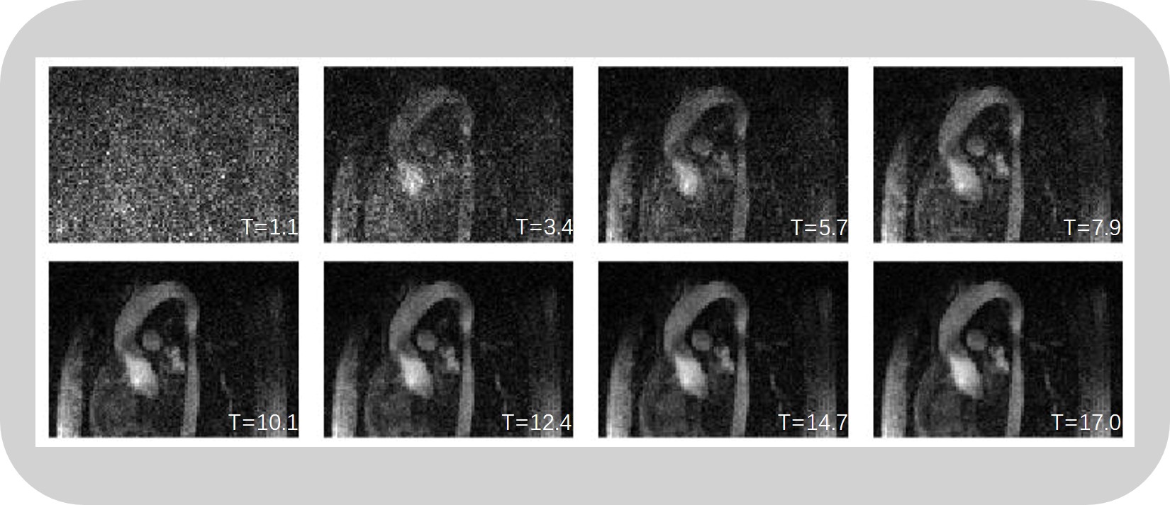
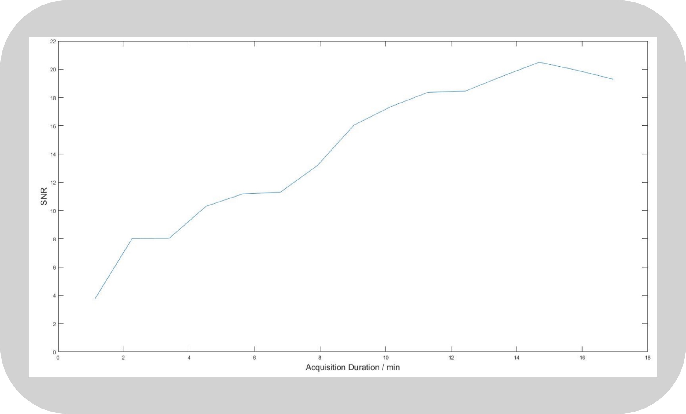
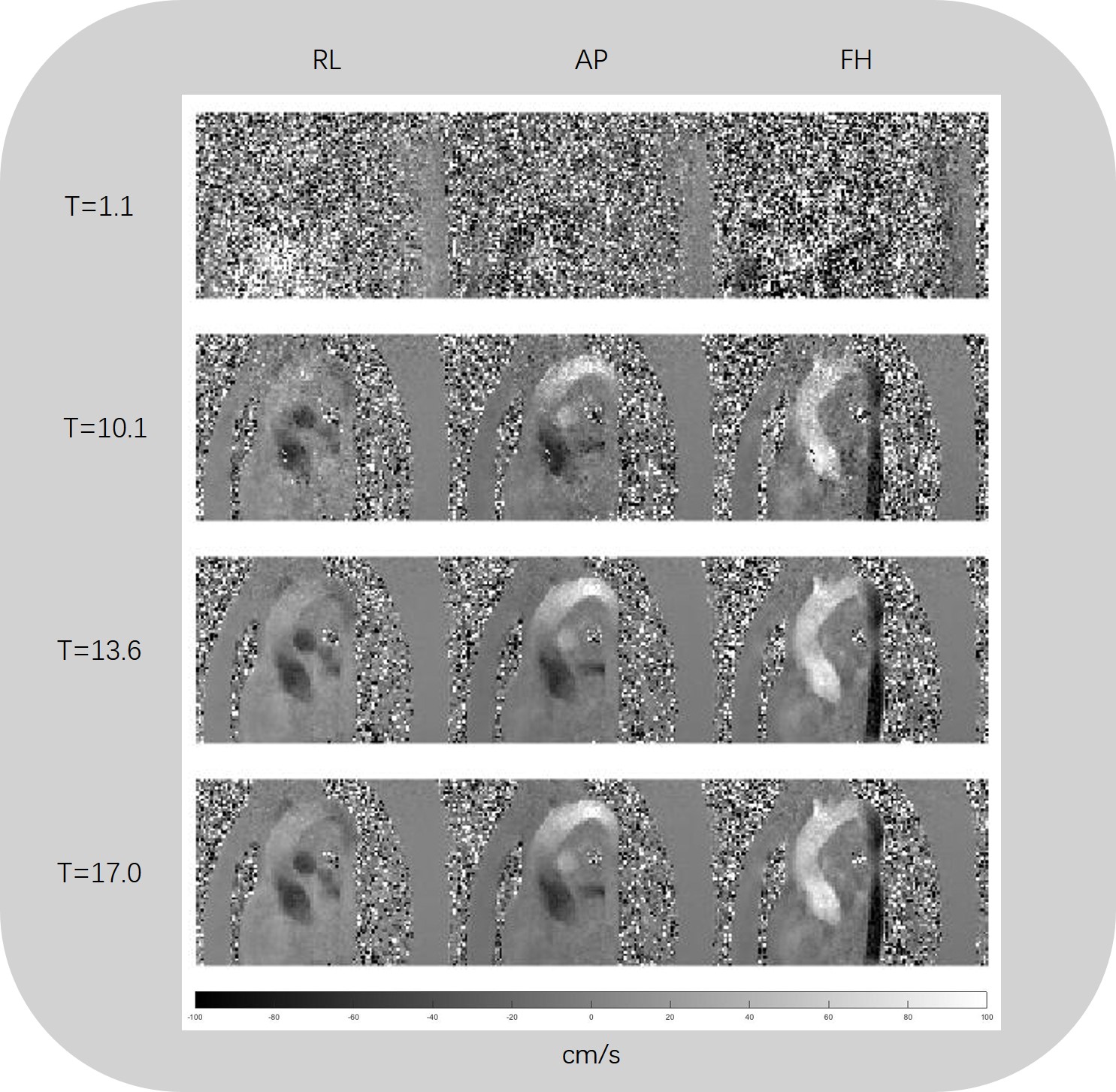
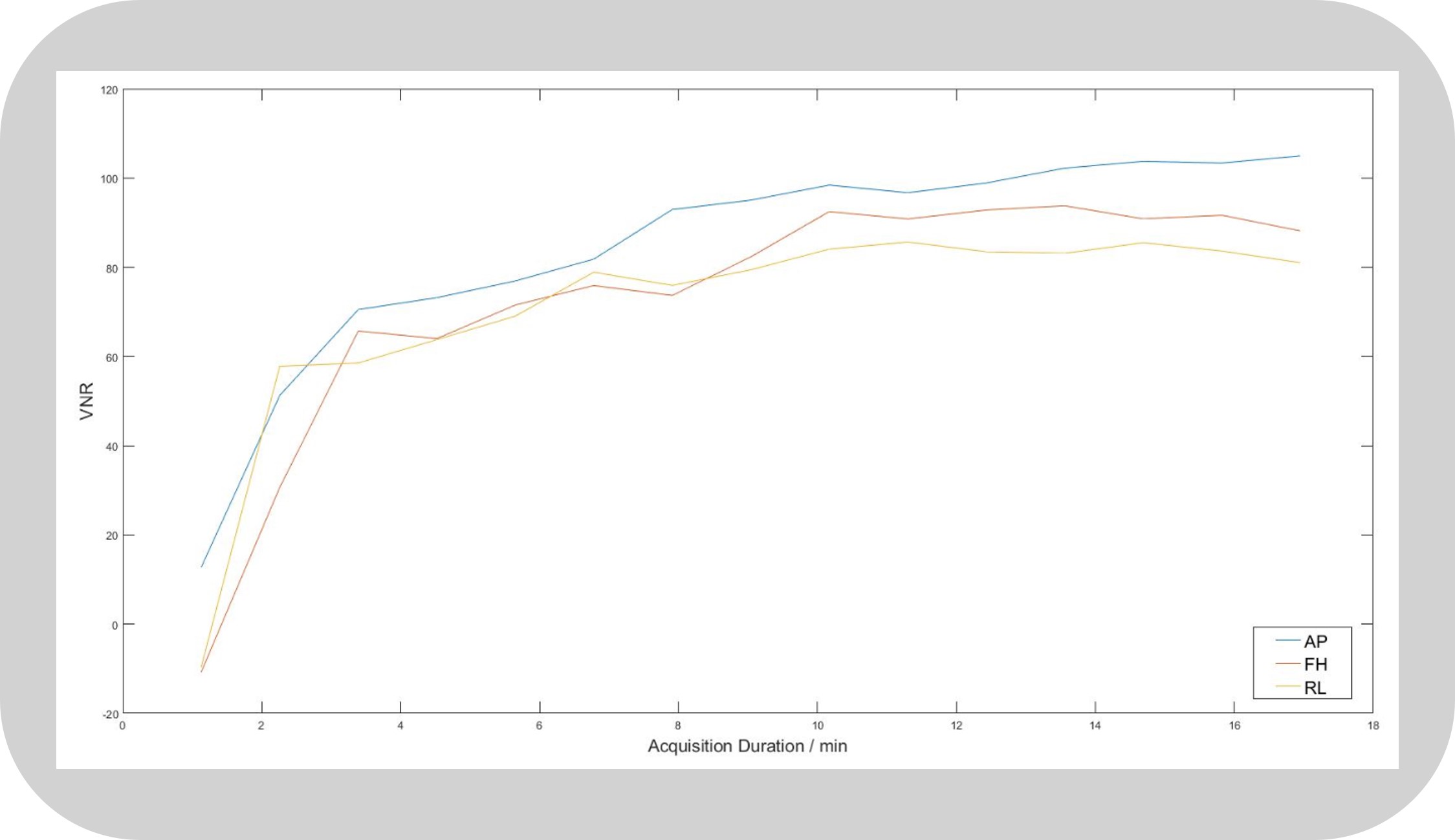
Reconstructed magnitude maps (in systole) at different acquisition duration were shown in Figure 1. Figure 2 showed the SNR curve with the increase of acquisition duration. When the acquisition duration was short, for example, T = 1.1min, the SNR was very low, and we can’t even distinguish the boundary of aorta. With the increase of acquisition duration, SNR increased rapidly. However, when the acquisition duration was long enough, for example, T > 13.6min, the SNR was converged without signification increase. Figure 3 showed the velocity maps (in systole) at different acquisition duration, and the VNR curves for three directions were shown in Figure 4. Similar to the magnitude maps and SNR curve, with the growth of acquisition duration, VNRs for three directions grew quickly and basically stayed at high values without big change. Meanwhile, the noise and artifacts for velocity maps gradually decreased and velocity maps gained good qualities at long acquisition duration (T > 13.6min).Discussion
We investigated the relationship between image
quality and acquisition duration for 4D flow MRI in this study. When the
acquisition duration is too short, the low-rank modeling suffers from the ill-conditioned
problems with small number of data. As a result, magnitude and velocity maps were
contaminated by noise and artifacts. With the increase of acquisition time, the
image quality improved and converged at 13.6 minutes in aorta scan. Further study
need to be performed to investigate the optimized acquisition duration for
different acquisition duration containing various information in both spatial
and temporal domains.Conclusion
In this study, we reconstructed the data from arbitrary acquisition duration for real-time 4D flow MRI, and assessed the image quality with different amount of data associated with acquisition duration for the low-rank modeling based real-time 4D flow MRI, which can provide an optimized scan time for real-time 4D flow MRI.Acknowledgements
No acknowledgement found.References
[1] Sun A, Zhao B, Li Y, et al. Real-time phase-contrast flow cardiovascular magnetic resonance with low-rank modeling and parallel imaging[J]. Journal of Cardiovascular Magnetic Resonance, 2017, 19(1): 19.
[2] Sun A, Zhao B, Li R, and Yuan C. High-Resolution 4D real-time phase-contrast flow MRI with sparse sampling. In Proceedings of the 25th Annual Meeting of ISMRM, Honolulu, Hawaii, 2017. p. 1261.
[3] Ringgaard S, Oyre S A, Pedersen E M. Arterial MR Imaging Phase-Contrast Flow Measurement: Improvements with Varying Velocity Sensitivity during Cardiac Cycle 1. Radiology, 2004, 232(1): 289-294.
Figures