2815
Effect of compressed sensing acceleration on high spectral and spatial resolution (HiSS) breast MRI image quality1Radiology, University of Chicago, Chicago, IL, United States, 2Fraunhofer MEVIS, Bremen, Germany
Synopsis
Strong T2* weighting has allowed high sensitivity of HiSS breast MRI to cancer, but in whole-breast imaging, contrast is compromised due to necessarily shorter echo trains. k-space under-sampling techniques such as compressed sensing (CS) yield time savings that can be traded for longer echo trains and stronger T2* weighting, potentially increasing breast HiSS MRI performance in screening and diagnostic applications. Our CS simulation resulted in minimal reduction in spatial resolution for acceleration factor R = 2, showing CS to be a promising acceleration strategy for HiSS MRI, allowing longer echo trains and stronger T2* weighting.
INTRODUCTION
Because of the risks of gadolinium contrast use, large MRI breast cancer screening programs are not viable without effective non-contrast enhanced MR sequences. EPSI-based High Spectral and Spatial resolution (HiSS) breast MRI has high diagnostic utility for lesion characterization, and could also potentially serve as a powerful tool for non-contrast enhanced breast cancer screening. [1, 2] However, whole breast HiSS MRI is currently implemented with short echo trains to reduce run time; this potentially reduces sensitivity. Compressed sensing (CS) acceleration reduces the number of phase encoding steps per slice, producing significant time savings which in HiSS MRI could be traded for longer echo trains. This would restore T2* weighting and higher spectral resolution that could facilitate effective breast cancer screening and diagnostic applications. Here, we demonstrate the application of CS to in vivo breast HiSS imaging, and evaluate its effect on image quality.METHODS
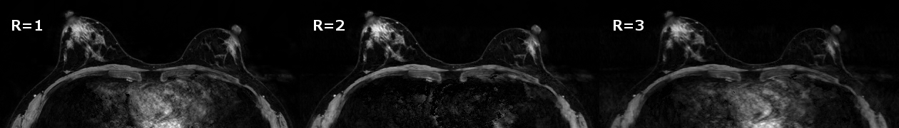
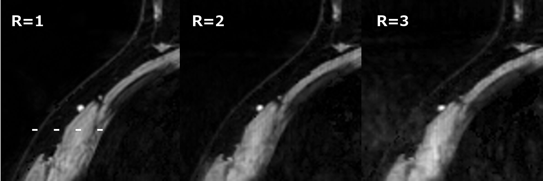
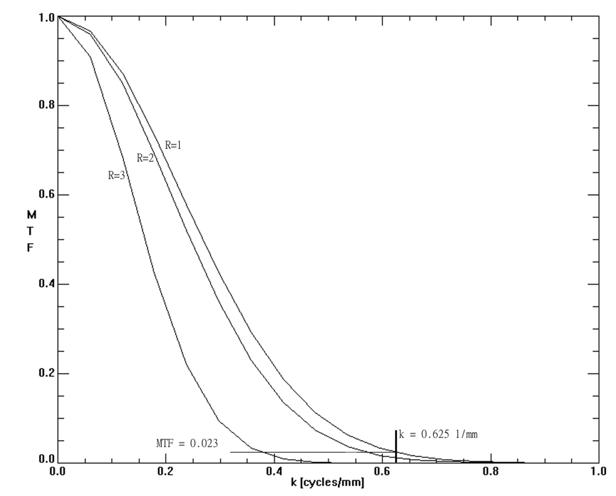
Whole-breast HiSS MRI was acquired on 1 patient on a Philips Achieva 3T-TX scanner, (2D multislice, 384 mm FOV, 0.8x0.8x3 mm3 voxels in 60 slices, TR/TE 2350/23 ms, 23 echoes, 23.9 Hz spectral resolution, scan time 7.5 min, SENSE factor 3 (L/R)). Because of SENSE reconstruction, k-space data was not directly available; instead, k-space data were calculated from reconstructed complex gradient echo images. CS acceleration was simulated by under-sampling the full k-space dataset, with three acceleration factors R. For R = 1, full k-space information was retained. For R = 2: variable-density random under-sampling, by a factor of 2 (pattern did not vary from echo to echo) was applied, and for R = 3 the same algorithm was applied, but using a factor of 3 under-sampling. After retrospective under-sampling, full k-space information was reconstructed for R = 2 and R = 3 using CS with sparsifying operators in spatial total variation and wavelet domains. [1] Complex gradient echo images, preserving phase information along the echo train, were reconstructed for R = 2 and 3. The complex gradient echo images were Fourier transformed along the temporal direction in order to obtain proton spectra in each individual voxel. The spectra resolved water and fat peaks, which were fit to Lorentzian functions in order to extract the pure water signal above the baseline and any lipid peak tails. [2] The water peak information thus obtained was used to generate water peak height images, with excellent fat suppression. [3] In order to quantify loss of image resolution with application of CS algorithm, signal profiles across the lateral or frontal edge of the chest wall muscle were extracted, to approximate a sharp edge. The profiles were positioned to be parallel to the readout and phase encoding directions and perpendicular to the chest wall. The profiles’ derivatives were fit to a Gaussian function which, after a Fourier transform, provided the modulation transfer function (MTF) for datasets with R = 1, 2, and 3. The loss of image resolution in both the readout and phase encoding direction was evaluated by comparing k values for an equal MTF level, with R = 1 dataset and k = 0.625 1/mm (equivalent to 0.8 mm resolution) used as a baseline.RESULTS
R = 2 and R = 3 datasets resulted in an effective spatial resolution in the phase-encoding direction of 0.88 and 1.33 mm, respectively, which compares to 0.8 mm for R = 1. The effective spatial resolution in the readout direction was 0.87 and 0.90, for R = 2, and 3, respectively.DISCUSSION
Our results indicate that minimal blurring due to CS implementation can be expected with two-fold acceleration. This would allow significant acceleration without correspondingly increased image quality loss. The sensitivity of HiSS MRI to breast lesions could be increased to allow non-contrast enhanced screening and diagnostic applications.CONCLUSION
CS acceleration simulation in in vivo breast HiSS MRI resulted in minimal reduction in spatial resolution for an acceleration factor of R = 2. Therefore, CS is a promising acceleration strategy for HiSS MRI. Faster HiSS acquisitions with longer echo trains and increased T2* weighting could improve sensitivity of HiSS breast imaging to breast lesions, and facilitate screening and diagnostic applications without the need to inject contrast media.Acknowledgements
No acknowledgement found.References
1. Lustig M, Donoho D, Pauly JM. Sparse MRI: The application of compressed sensing for rapid MR imaging. Magnetic resonance in medicine 2007; 58:1182-1195
2. Medved M, Ivancevic MK, Olopade OI, Newstead GM, Karczmar GS. Echo-planar spectroscopic imaging (EPSI) of the water resonance structure in human breast using sensitivity encoding (SENSE). Magnetic resonance in medicine 2010; 63:1557-1563
3. Fan X, Abe H, Medved M, et al. Fat suppression with spectrally selective inversion vs. high spectral and spatial resolution MRI of breast lesions: qualitative and quantitative comparisons. Journal of magnetic resonance imaging : JMRI 2006; 24:1311-1315
4. Medved M, Fan X, Abe H, et al. Non-contrast enhanced MRI for evaluation of breast lesions: comparison of non-contrast enhanced high spectral and spatial resolution (HiSS) images versus contrast enhanced fat-suppressed images. Academic radiology 2011; 18:1467-1474
Figures