2805
Real-time cardiac cine using supervised machine learning and compressed sensing with radial trajectory1UIH America., Houston, TX, United States
Synopsis
2D Real-time cardiac cine imaging is valuable for myocardiac function studies. Compared with Cartesian trajectory, Golden-angle (GA) radial acquisition is promising in patients with impaired breath-hold capacity [1]. The GA radial acquisition is an easy-to-implement and promising technique that features improved spatial-temporal resolution, and overcuts Cartesian sampling trajectories in reducing motion artifacts.
Introduction
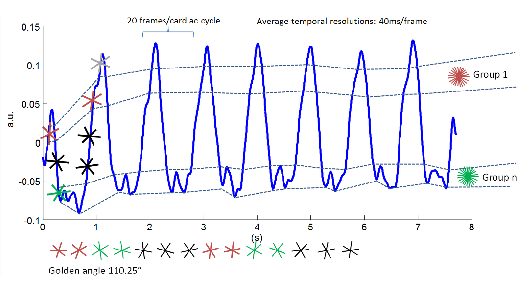
The need for ECG gating presents many difficulties in cardiac magnetic resonance imaging. ECG free cardiac cine imaging using golden-angle (111.25°) radial acquisition is desirable in that it achieves relatively uniform angular distribution of radial spokes, therefore permits arbitrary choice of temporal resolution. Yet, existing reconstruction methods for real time radial cardiac MRI includes K-t RASPS [2], which is based on NUFFT [3] and SPARSE SENSE [4]; and the partial separability method [5], which enables real-time cardiac MRI without triggering, gating, or breath holding, but requires an accurate temporal basis. The temporal basis is usually calculated from fixed k-space locations at different temporal frames, which make it difficult to apply on radial sampling trajectory patterns. This abstract presents a new method to reconstruct cardiac cine with high spatial-temporal resolution using supervised learning, low rank model, parallel imaging, and compressed sensing. In data acquisition, k-space data were continuously acquired using a 2D radial bSSFP sequence with spoke angles incremented by the golden-angle. In reconstruction, the spokes were first grouped to provide enough k-space data for initialization, using k-nearest neighbor (k-NN) algorithm. Low rank model, compressed sensing, and Multi-channel Blind deconvolution (MalBEC) [6] were jointly used to reconstruct the final real-time cine images.Theory and methods
The main goal is to recover real time cardiac cine images \[{\Gamma ^{M \times N}} = [\gamma (x,{t_1}), \cdots ,\gamma (x,{t_N})]\] from its highly under-sampled radial spokes acquired through multiple channels of a phased array coil, as well as post-artificially gated cardiac cine images $$$\gamma (x,n)$$$ , where $$$x$$$ represents spatial location, $$$t$$$ is the time, and $$$n$$$ counts all cardiac phases.
Model: The image reconstruction problem is formulated as $$$\mathop {\min }\limits_{{\Gamma ^{M \times N}}} \sum\nolimits_{\ell = 1}^{Nc} {||{d_\ell } - {F_{\Omega ,\ell }}{\Gamma ^{M \times N}})||_2^2} + {\lambda _1}\sum\nolimits_{t = {t_1}}^{{t_N}} {||\gamma (x,t) - \gamma (x,n(t))||_2^2} + {\lambda _2}||{\rm{vec}}({\Gamma ^{M \times N}}{{\bf{F}}_t})|{|_1}$$$
where the first term is the data consistency term, $$${d_\ell }$$$ represents the acquired spokes from the $$${\ell }$$$-th channel, $$${F_{\Omega ,\ell }}$$$ is an operator which integrates both the Fourier transform with a specified undersampling trajectory in (k,t)-space and the coil sensitivity modulation; the second term regularizes real time cardiac cine images corresponding to the post-artificially gated ones; the L1 norm term enforces the sparseness in spatial and temporal frequency domain, $$${{\bf{F}}_t}$$$ represents the temporal Fourier transform. The k-NN is a non-parametric method used for classification and regression. In this work, k-NN method was used to cluster the spokes. The center point values (all channels) of all radial spokes were used to cluster these spokes into R groups. We assume these center point values present low rankness. As a result, R can be small. For each group, the corresponding post-artificially gated cine images $$$\gamma (x,n)$$$ were reconstructed from MalBEC.
Results
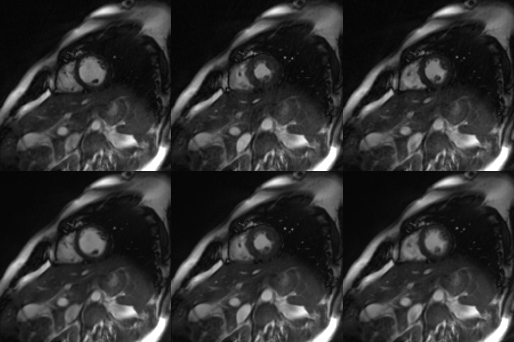
The proposed method was implemented on a research 1.5T scanner (uMR560, United Imaging Healthcare, Shanghai, China). Imaging parameters were: TE/TR =1.46/2.92 ms, bandwidth = 1000 Hz, spatial resolution = 1.9x1.9 mm2, FOV = 360x360 mm2, slice thickness = 10 mm, flip angle =80°. A fixed number of 2000 spokes were acquired regardless of the heart rate.Conclusion
We have proposed an approach to reconstruct real-time cardiac cine from radial samplings without ECG signal. The approach first reconstructed artificially-gated cine images using supervised machine learning and MalBEC. Based on the gated cine images, real-time images were also reconstructed with only 15 spokes per frame, corresponding to an average temporal resolution of 40 ms per frame.
Acknowledgements
No acknowledgement found.References
[1] P Kellman, et al., MRM v.62, pp.1557, 2009.
[2] L Feng, et al., ISMRM, pp. 225, 2005.
[3] JA Fessler. IEEE T-SP 2003 51(2):560-74.
[4] R Otazo, et al. MRM 2010; 64:767-776.
[5] C Brinegar, et al., Conf Proc IEEE Eng Med Biol Soc., pp.3381–3384, 2008.
[6] J Lyu, et al., ISMRM, pp. 3232, 2016.
Figures