2725
Quantitative texture analysis of apparent diffusion coefficient (ADC) for evaluating histologic differentiated grade of head and neck squamous cell carcinomaYu Chen1, Yanan Zhao1, Huadan Xue1, Zhuhua Zhang1, and Zhengyu Jin1
1Peking Union Medical College Hospital, Beijing, China
Synopsis
To investigate the feasibility of using texture analysis (TA) of apparent diffusion coefficient (ADC) to distinguish between well- and moderate- differentiated head and neck squamous cell carcinoma (HNSCC). A total of 22 patients were retrospectively analyzed, including: well-differentiated degree SCC (WSCC, n=11) and moderate-differentiated degree SCC (MSCC, n=11). A Mean>101.38 at coarse texture scale (SSF=6mm) identified WSCC and MSCC with the highest AUC of 0.843±0.083 (Se=72.7%, Sp=81.8%, PPV=80%, PV=75%, and accuracy=77.3%). Texture analysis of ADC proved to be a feasible tool for differentiating WSCC from MSCC, and had better diagnostic performance than ADC value.
Introduction/purpose
The histopathological classification of malignancy proposed by the World Health Classification (WHO, 2005) was based on the degree of cell differentiation and allowed the classification of this malignancy into three categories, as well-, moderately and poorly differentiated. This is increasingly challenged by new non-invasive advanced MRI techniques and research into additional sequences to improve radiological diagnostic accuracy. Texture analysis (TA) assessed the distribution of gray-levels within an image to obtain texture features of intra-lesional heterogeneity. The aim of our study is to investigate the feasibility of using texture analysis (TA) of apparent diffusion coefficient (ADC) to distinguish between well- and moderate- differentiated head and neck squamous cell carcinoma (HNSCC).Methods
A total of 22 patients undergoing MR examination with diffusion weighted image (DWI) before treatment were retrospectively analyzed. All patients were histological proven as HNSCC, including: well-differentiated degree SCC (WSCC, n=11) and moderate-differentiated degree SCC (MSCC, n=11). The ADC mapping was derived from DWI on workstation. The minimum, average and maximum ADC values were compared between the WSCC and MSCC. The largest cross-section area of the tumors were chosen for texture analysis using TexRAD software. Comparing of texture parameters, mean gray-level intensity (Mean), standard deviation, entropy, mean of positive pixels (MPP), skewness, and kurtosis were made for the objective. Receiver operating characteristic (ROC) analysis was performed and the area under the ROC curve was calculated for texture parameters that were significantly different (P<0.05) for the purpose. Sensitivity (Se), specificity (Sp), positive predictive value, negative predictive value, and accuracy were calculated using the cut-off value of texture parameters with the highest AUC.Results
The minimum, average and maximum ADC values showed no significant difference between WSCC and MSCC. Compared to MSCC, WSCC had significantly higher Mean from fine, medium and coarse texture scale (P<0.05), higher SD from non-filtration and fine scale (P<0.05), higher MPP from fine and medium scale (P<0.05), and lower entropy from all scale (P<0.05). There was no significant difference in skewness or kurtosis at any texture scale of ADC. A Mean>101.38 at coarse texture scale (SSF=6mm) identified WSCC and MSCC with the highest AUC of 0.843±0.083 (Se=72.7%, Sp=81.8%, PPV=80%, PV=75%, and accuracy=77.3%).Discussion
This study demonstrated the potential for TA of ADC to distinguish between the WSCC and MSCC by quantifying heterogeneity, without additional imaging. TA is an easy post-processing step that can be performed on existing DICOM format images. The results can in part be explained by the correlation between tumour heterogeneity and tumour grade.Conclusions
Texture analysis of ADC proved to be a feasible tool for differentiating WSCC from MSCC, and had better diagnostic performance than ADC value. Mean quantified from coarse texture scale was the optimal diagnostic parameter for estimating histologic differentiated degree of HNSCC.Acknowledgements
Thank you for the support from Professor Balaji Ganeshan, Institute of Nuclear Medicine, University College London, University College Hospital.References
References [1] Schob S, Meyer HJ, Dieckow J, et al. Histogram Analysis of Diffusion Weighted Imaging at 3T is Useful for Prediction of Lymphatic Metastatic Spread, Proliferative Activity, and Cellularity in Thyroid Cancer. Int J Mol Sci. 2017. 18(4). [2] Carrillo JF, Carrillo LC, Cano A, et al. Retrospective cohort study of prognostic factors in patients with oral cavity and oropharyngeal squamous cell carcinoma. Head Neck. 2016. 38(4): 536-41. [3] Surov A, Stumpp P, Meyer HJ, et al. Simultaneous (18)F-FDG-PET/MRI: Associations between diffusion, glucose metabolism and histopathological parameters in patients with head and neck squamous cell carcinoma. Oral Oncol. 2016. 58: 14-20. [4] Skogen K, Schulz A, Dormagen JB, Ganeshan B, Helseth E, Server A. Diagnostic performance of texture analysis on MRI in grading cerebral gliomas. Eur J Radiol. 2016. 85(4): 824-9.Figures
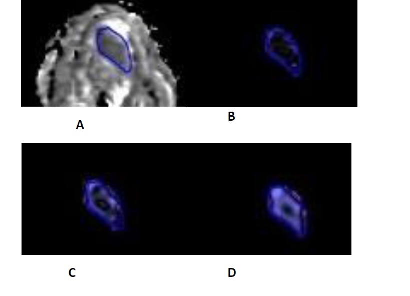
A example
of texture analysis of ADC map. A 60 years old, male, with tongue cancer..The histological results demonstrated it was well-differentiated degree. The
lesion showed lower ADC value on the ADC map (A). The texture analysis showed
different heterogeneity in fine(B), moderate (C) and coarse (D) filtration
scale.