2708
Simultaneous Multi-Slice fMRI of the Mouse Brain Using POMP-EPI at 9.4T1Queensland Brain Institute, The University of Queensland, St Lucia, Australia, 2Centre for Advanced Imaging, The University of Queensland, St Lucia, Australia
Synopsis
Acceleration of rodent brain functional MRI using parallel imaging techniques is not widely used due to the limited availability of high-density phased-array coil on pre-clinical scanners. In this study we demonstrated a POMP-EPI method to enable simultaneous multi-slice acquisition for fast mouse brain imaging without a phased array coil. A four-fold multiband acceleration was achieved without using coil sensitivity information. This method can be used to increase the spatial or temporal resolution of mouse fMRI acquisition, which will benefit the study of dynamics of neural activity and connectivity.
INTRODUCTION
In recent
years simultaneous multi-slice EPI (SMS-EPI) has been widely adopted in human
brain imaging to achieve high spatial and temporal resolution for fMRI and DTI acquisitions1,2.
The SMS-EPI technique excites multiple slices simultaneously, and uses parallel
imaging methods to reconstruct individual slice images from the aliased and
mixed data. As parallel imaging reconstruction requires large number of
receiving coils in the accelerated direction to provide necessary spatial information,
it is currently difficult to apply SMS techniques to rodent brain imaging because
of the limited availability of proper receiving coil arrays on preclinical MRI
system.
POMP-EPI3 uses blipped-CAIPI method4 that utilizes blipped gradient
along the slice-select direction to shift multi-slice images to different
phase-encoding positions and removes aliasing. It was demonstrated in human
brain functional imaging and allows acceleration in slice-select direction
without the need for multiple receiving channels3.
In this
study we implemented POMP-EPI sequence for mouse brain imaging at 9.4T. With a multiband factor
of 4, a TR of 300ms was achieved for covering the whole brain with 0.3x0.3x0.6
mm3 spatial resolution.METHODS
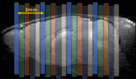
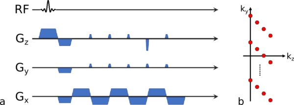
Figure 1 shows the POMP-EPI slice acquisition scheme. A RF pulse with 4 bands that are 3mm apart was used for excitation. Slice select gradient blips were applied between the readout lines to introduce slice-dependent linear phase shifts that push the slices FOV/4 apart from each other, so the aliasing can be avoided with increased FOV (Figure 2). Mice cadaver were scanned on a 9.4T pre-clinical scanner (Biospec 9.4/30, AVIII HD, PV6.0.1, Bruker BioSpin MRI GmbH, Ettlingen, Germany) with a Tx/Rx cryoprobe. Conventional gradient-echo EPI contains 20 single-slice excitations per TR, where slice thickness was 0.5mm with 0.1mm gaps. TR/TE = 1000/10.8 ms, α = 60°, FOV = 19.2x19.2mm2, matrix size = 64x64 (in-plane resolution = 0.3x0.3 mm2) was acquired for 360 repetitions (6 min). POMP-EPI scans had an extended FOV of 19.2x38.4mm2 while keeping the same spatial resolution. A 4-fold multiband acceleration reduced the repetition time to 300 ms and α = 30° but increased TE to 17.6 ms for full k-space acquisition (matrix size = 64x128) which can be reduced to 10.8 ms by partial Fourier acquisition (PF-POMP-EPI, matrix size = 64x96). Partial Fourier data were reconstructed by phase-constrained POCS method5.RESULTS AND DISCUSSION
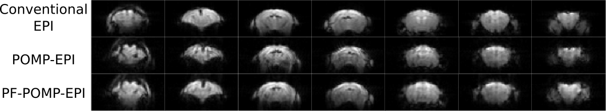
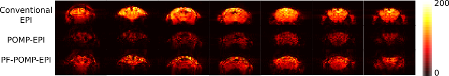
Figure 3 shows example slices from each imaging sequence. POMP-EPI has prolonged TE and acquisition window, which led to more signal decay, susceptibility artifacts and distortion. The artifacts were partially reduced by the use of PF-POMP-EPI. Correction based on reversed-phase acquisition and estimated field coefficient could be used to further reduce the distortion. Figure 4 shows the tSNR of example slices of each sequence over a 6-minute scan. The tSNR of POMP-EPI and PF-POMP-EPI were lower (mean tSNR = 20.5 And 31.4, respectively) compared to conventional EPI (tSNR = 70.3) due to the much shorter TR and lower flip angle. However the substantially increased number of time points (3.3x in this study) for the same scan duration could effectively compensate for the detection power in an fMRI experiment.CONCLUSION
We implemented POMP-EPI sequence on 9.4T scanner for fast mouse whole-brain imaging without the use of high-density receiving coil array. A temporal resolution of 300ms could be achieved while maintaining the same spatial resolution and coverage. The increased speed can be used to attain higher degrees of freedom for the fMRI time-series or to further increase the through-plane spatial resolution with more slices. This method could also be extended to spin-echo EPI for accelerating diffusion tensor imaging.Acknowledgements
The authors thank the Centre for Advanced Imaging at the University of Queensland for imaging support.References
1Moeller S, Yacoub E, Olman CA, et al. Multiband multislice GE-EPI at 7 Tesla, with 16-fold acceleration using partial parallel imaging with application to high spatial and temporal whole-brain fMRI. Magn Reson Med 2010;63:1144–1153.
2Feinberg DA, Moeller S, Smith SM, et al. Multiplexed echo planar imaging for sub-second whole brain FMRI and fast diffusion imaging. PLoS One 2010;5:e15710.
3Lan P, Islam H, Glover G. Phase-offset Multiplanar Echo-planar Imaging (POMP-EPI) to Accelerate fMRI Acquisition without Parallel Imaging. Proc. ISMRM 2017, #1632
4Setsompop K, Gagoski BA, Polimeni JR, et al. Blipped-Controlled Aliasing in Parallel Imaging for Simultaneous Multislice Echo Planar Imaging with Reduced g-Factor Penalty. Magn Reson Med. 2012; 67:1210–1224.
5Liang Z- P, Boada FE, Constable RT, et al. Constrained reconstruction methods in MR imaging. Reviews of Magn Reson Med. 1992; 4:67–185.
Figures