2702
Respiratory-Gated B0 Field Stabilisation for High Resolution Mouse Brain Imaging1CRUK/MRC Oxford Institute for Radiation Oncology, University of Oxford, Oxford, United Kingdom
Synopsis
The echoes of a 3D multi gradient echo (MGE) scan are typically combined for detection of USPIO and MPIO. The echo combination requires B0 to be constant throughout the scan to achieve good image fidelity at high resolution. A navigator acquisition embedded in the MGE scan maintains the MR steady state and enables a real-time adaptive B0 correction. It is demonstrated that a respiratory-gated correction scheme outperforms ungated correction in mouse brain for the detection of micron sized iron-oxide particles coupled with anti-vascular cell adhesion molecule antibody (VCAM-MPIO) to identify inflammation in vessels.
Introduction
3D multi gradient echo (MGE) enables efficient high-resolution scanning with T2* contrast for the detection of USPIO and MPIO. Echoes are often combined to generate an image with improved SNR from the early echoes and boosted contrast from later echoes as in the MEDIC/MERGE/M-FFE/ADAGE methods.1-3 However, a useful combined image is only obtained if the image of each echo is properly aligned, which requires B0 to be constant throughout the scan.
Rapid scanning causes gradient heating, which induces a B0 drift that predominantly affects image registration in the slow phase-encoding dimension of 3D MGE images. For sequential phase-encode order this applies an additional phase ramp to the raw data, the magnitude of which is proportional to TE, such that successive echoes appear shifted and distorted in image space.
A real-time adaptive B0 correction during 3D MGE scanning has been developed to reduce this effect. The respiratory-gated B0 correction is demonstrated to outperform ungated B0 correction and improves image fidelity.
Methods
MRI was performed on a 7.0 T, 210 mm horizontal bore scanner equipped with 120 mm bore gradient insert (Varian) and a 30 mm long 25 mm ID quadrature birdcage coil (Rapid Biomedical). The mouse head was immobilised with a headframe and respiration was monitored using a pressure balloon.
3D MGE was performed with TR 50 ms, TE 3 ms, NE 12, ESP 3.5 ms, FA 12º, FOV 22.5×22.5×22.5 mm3, and matrix 256×192×192 zero-filled to 256×256×256 to give 88 µm isotropic resolution in a scan time of 30.9 mins.
A navigator acquisition evaluation with B0 update was inserted throughout the scan as shown in Figure 1. A TTL controlled switch routed the NMR signal to the scanner ADC or a B0 correction unit, which mixed the signal to approximately 1000 Hz for frequency measurement using a microcontroller (Arduino). The measured frequency was used to generate a calibrated voltage across the B0 correction coil that was combined with the scanner’s real-time B0 correction to maintain the measured frequency offset at 1000 Hz. Scans were operated with and without respiratory-gated conditional B0 update.
Characterisation of the effects of B0 drifts and validations of B0 corrections were performed in normal mice (n=6). Acute brain inflammation was induced by unilateral stereotactic injection of interleukin 1β (IL-1β) into the left striatum of BALB/c mice (n=2). Animals were injected intravenously with micron sized iron-oxide particles coupled with anti-vascular cell adhesion molecule antibody (VCAM-MPIO) to detect inflamed vessels in the IL-1β injected hemisphere of the brain.4
Anaesthesia was induced with 4% isoflurane in air and maintained with 1-3% isoflurane in a 1:5 O2:air mixture for surgery and MRI to give a respiration rate of 40-60 breaths/minute. Rectal temperature was maintained at 35-37 °C.
To assess image fidelity, data were automatically analysed for hypointense voxels in the brain, as for MPIO quantification.5
Results
Figure 2 shows that the TE dependent image corruption observed without B0 correction was significantly reduced with respiratory-gated B0 correction.
Figure 3 shows the B0 measurements from 12 s bursts of navigator signal acquisitions that were acquired every 10 minutes during operation of an uncorrected MGE scan. Rapid frequency changes resulting directly from respiratory motions are superimposed upon a longer-term frequency drift caused by gradient induced heating.
The stability of the frequency measurement was improved when conditional, respiratory-gated measurements were made. Figure 4 shows frequency measurements calculated from the navigator signals during 3D MGE scanning with respiratory-gated and ungated B0 correction. The comparison shows that better frequency stability is achieved when both respiratory and gradient heating derived instabilities are minimised.
The most detailed images were produced when the MGE scan was operated in a stable B0. Figure 5 compares the efficacy of the B0 correction schemes with an uncorrected scan in three sequential scans, throughout which the system slowly approaches (but never reaches) thermal equilibrium.
The image acquired with respiratory-gated B0 correction shows improved detail of MPIO, whilst the image acquired with ungated B0 correction exhibits ghosting from the jitter in frequency measurement caused by respiration. In the uncorrected image a 27 Hz shift during scanning results in a loss of image fidelity with respect to the respiratory-gated B0 corrected scan.
The improved image fidelity of respiratory-gated and ungated B0 correction increased the hypointense brain voxel count measured in uncorrected data by 38% and 24%, respectively.
Conclusion
Gradient heating and respiratory sources of B0 field instability lead to image corruption that can be reduced with a respiratory-gated B0 correction scheme. The improved fidelity of MGE imaging of mouse brain incurs only a 3% time penalty.
Acknowledgements
The work was supported financially by Cancer Research UK (CRUK grants C5255/A12678, C2522/A10339), the Engineering and Physical Sciences Research Council (EPSRC grant C2522/A10339) and the Medical Research Council Unit Grant for the Oxford Institute for Radiation Oncology.References
1. Ward J, Guthrie JA, Wilson D, Arnold P, Lodge JP, Toogood GJ, Wyatt JI, Robinson PJ. Colorectal hepatic metastases: detection with SPIO-enhanced breath-hold MR imaging–comparison of optimized sequences. Radiology 2003;228:709-18.
2. Choi JS, Kim MJ, Kim JH, Choi JY, Chung YE, Park MS, Kim KW. Comparison of multi-echo and single-echo gradient-recalled echo sequences for SPIO-enhanced liver MRI at 3 T. Clin Radiol 2010;65:916-23.
3. Schieda N, Avruch L, Shabana WM, Malone SC. Multi-echo gradient recalled echo imaging of the pelvis for improved depiction of brachytherapy seeds and fiducial markers facilitating radiotherapy planning and treatment of prostatic carcinoma. J Magn Reson Imaging 2015;41(3):715-20.
4. McAteer MA, Sibson NR, von Zur Muhlen C, Schneider JE, Lowe AS, Warrick N, Channon KM, Anthony DC, Choudhury RP. In vivo magnetic resonance imaging of acute brain inflammation using microparticles of iron oxide. Nat Med. 2007;13(10):1253-8.
5. Serres S, Soto MS, Hamilton A, McAteer MA, Carbonell WS, Robson MD, Ansorge O, Khrapitchev A, Bristow C, Balathasan L, Weissensteiner T, Anthony DC, Choudhury RP, Muschel RJ, Sibson NR. Molecular MRI enables early and sensitive detection of brain metastases. Proc Natl Acad Sci U S A. 2012;109(17):6674-9.
Figures
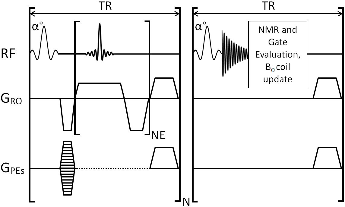
Figure 1. 3D MGE scan with embedded navigator acquisition that maintains the MR steady state. Inserting a navigator acquisition after every N=32 PE steps increases the scan time by less than 1 min. NMR signal frequency evaluation is used to adjust the voltage across the B0 coil during the same TR. A respiration gated B0 update is performed only if the NMR measurement occurs during a stable inter breath period.

Figure 2. 3D MGE echoes acquired with and without respiratory-gated B0 correction. The ROI overlay highlights the image movement in the slow PE dimension in the absence of B0 correction and good image alignment with respiratory-gated B0 correction. TR 50 ms, TE 2.75 ms, NE 16, ESP 2.75 ms, FA 12º, FOV 32×32×32 mm3, matrix 256×192×192 zero-filled to 256×256×256 to give 125 µm isotropic resolution in a scan time of 30.9 mins without correction and 31.8 mins with correction. Without B0 correction the image shift between Echoes 1 and 15 was calculated and measured to exceed 8 pixels (1 mm).

Figure 3. 12 s bursts of continuous navigator signal measurement at 10 min intervals during 3D MGE scanning. The respiration cycle of the mouse is clearly detectable within each burst and a long term drift results from gradient warming. The navigator signal is acquired 1000 Hz off-resonance so that positive and negative frequency shifts are easily distinguishable.

Figure 4. Frequency measurements calculated from the navigator signals during 3D MGE scanning for respiratory-gated and ungated scans. The navigator signal is acquired 1000 Hz off-resonance so that positive and negative frequency shifts are easily distinguishable. The measurement range was 17.7 Hz for the respiration gated correction and 36.8 Hz for the ungated correction.

Figure 5. Sum of squares combination image slices generated from the echoes of 3D MGE data of an inflammation model injected with VCAM-MPIO. Data were acquired with respiratory-gated B0 correction followed by ungated B0 correction followed by no correction. The frequency shift measured over the successive scans reduces as the system approaches thermal equilibrium. Although the respiratory-gated B0 corrected data were acquired during the greatest B0 instability the resulting images exhibit the best fidelity. The right hand side of each image display is ipsilateral to the interleukin 1β injection.